Functions of frontal fractions. How does the brain arranged: the frontal shares of the middle frontal convolution for what is responsible
The frontal shares are located in the front of the brain, in front of each cerebral hemisphere and in front of the parietal fraction. They are considered the most important department because of their functions and because they take one third of the total brain. In other species, their volume is lower (17% chimpanzee and a dog of 7%). They play a role in motion management, as well as in the mental functions of the highest level, behavior and emotional control.
Building and location
The frontal shares are divided into two main areas: motorcor and prefrontal bark. The brain area participating in the language and speech, known as Brock, is located in the left frontal share.
The prefrontal bark is the front of the frontal fraction and controls complex cognitive processes, such as memory, planning, reasoning and solving problems.
This frontal share area helps to establish and maintain targets, restrain negative impulses, organize events in temporary order and form individual identities.

Functions of frontal fractions
Lob's share regulates motivational processes. They are also responsible for the perception and resolution of conflicts, as well as for constant attention, for controlling emotions and social behavior. They regulate emotional processing and control the context-based behavior.
Functions of prime crust
The main function of the motor crust is to control arbitrary movement, including expressive, writing and eye movement. Primary motor core sends the neuron team to the brain barrel and spinal cord. These are responsible for specific voluntary movements. Inside the primary motor cortex of two hemispheres, there is a representation of a contralateral half of the body. That is, in each hemisphere there is a representation of the opposite side of the body. This area controls the programming of preparation and movement. Premotor Cora automates, harmonizes and archives movements related to previous experiments.
The primary motor bark of frontal fractions is involved in arbitrary movement. It has nervous ties with a spinal cord, which allow this area of \u200b\u200bthe brain to control muscle movements. Movement in various fields of the body is controlled by primary motorbate, each region is associated with a specific engine crust area. Parts of the body requiring fine motion control occupy large areas of the motor cortex, and those that require simpler movements occupy less space. For example, plots of motor cortex controlling the movement of the face, language and arms occupy more space than areas associated with hips and torso. Primorous bark of frontal stakes has nervous bonds with primary motor bark, spinal cord and brain barrel. Premotor bark allows you to plan and perform the correct movements in response to external signals. This cortical area helps to determine the specific direction of movement.
Functions of prefrontal crust
The prefortional bark is located in front of the frontal lobe. It is considered the final expression of the development of the human brain. She is responsible for knowledge, behavior and emotional activity. The prefrontal bark receives information from the limbic system (participates in emotional control) and acts as a mediator between knowledge and feelings through the executive functions. Executive functions are a set of cognitive skills necessary to control and self-regulating behavior.
Functions of the Dorsolateral region of prefrontal bark
This is one of the most recently formed parts of the human brain. It establishes ties with three other areas of the brain and transforms information into thoughts, solutions, plans and actions.
She is responsible for cognitive abilities, such as:
- attention;
- focus;
- braking;
- maintenance and processing of information;
- programming upcoming actions;
- analysis of possible results;
- self-analysis of cognitive activity;
- analysis of the situation and the development of an action plan;
- ability to adapt to new situations;
- organization of behavior in relation to the new goal.
Frontal shares and associated disorders
Lobal lobes are involved in different processes (cognitive, emotional, behavioral). That is why the lesions caused by injuries caused to this area can vary from the symptoms of the brain shock to others, more serious.
Damage with a frontal share can lead to a number of difficulties, such as loss of fine motor function, difficulties with speech and tongue, difficulty thinking, inability to understand humor, lack of expression of the person and personality change.
Damage of the frontal share can also lead to dementia, memory disorders and the absence of impulse control.
Types and features of disorders in injuries
Damage to the primary or prime cortex can cause difficulties in coordinating the speed, execution and motion, which leads to different types of aprage. - This is a disorder in which a person has difficulty planning to perform tasks, provided that the request or command is clear and he / she wishes to perform the task. Ideasotor apraxia is a deficit or difficulty in the ability to plan or perform previously studied motor actions, especially those that need tool. The affected people can explain how to perform actions, but cannot act. Kinetic apraxia: voluntary limb movements are violated. For example, people cannot use their fingers in a coordinated way (playing piano). In addition to apraxia, other disorders can develop from a frontal share injury, such as language disorders or aphasias. Transcortic motor AFAZIO: Language disorder, due to a person there is no verbal fluency (slow-down speech and poorly organized), a limited spontaneous language (lack of initiative) and difficulties or disability in the letter. Brock: Language disorder, which generates a lack of verbal fluency, anomy (inability to access vocabulary to name words), poor syntax design in speech, recurrent, reading and writing difficulties. However, the symptoms will depend on the damaged area.

Dorsolateral region and injury
Injury in this area is usually associated with such cognitive problems as:
- The inability to solve complex problems: reducing the level of flexibility (reasoning, adaptation and solution of new situations, etc.).
- Cognitive rigidity and perseverance: a person supports thought or action, despite the proposal to change the thought or action.
- Reduced learning ability: difficulties in acquiring and maintaining new information.
- Memory disorder.
- Deficit in programming and change of motor activity: difficulties in organizing a sequence of movements and a change in activity.
- Reducing verbal flowability: deterioration of the ability to memorize words. This action requires not only the lexical part, but also the organization, planning, focusing and electoral attention.
- Acting attention: It is difficult to maintain attention and impede other irrelevant incentives or change focus of attention.
- Pseudodepressive disorders: Symptoms of depression (sadness, apathy, etc.).
- Reducing spontaneous activity, loss of initiative and motivation: noticeable apathy.
- : It is difficult to identify emotions and, therefore, the inability to express our own emotions.
- Language limitation: Answers are usually one-room.
Orbital region and injury
Symptoms of injury in this area are more behavioral. The behavior of a person tends to dissent (just what happened to the Phineas Gage, survived negative personal changes after the head injury):
- Irritability and aggressiveness: exaggerated emotional reactions in everyday life.
- Echopraxia: imitation of observed movements.
- Termination and impulsiveness: no self-control over behavior.
- The difficulty of adapting to social standards and rules: socially unacceptable behavior.
- Violation of judgment.
- Lack of empathy: difficulty understanding of other people's senses.
The frontal shares are incredibly important in order for people to be in force. Even without injuries of the brain, it is extremely important to maintain informative skills active - the brain health is important for full-fledged life.
The frontal share occupies the front semi-guns. From a parosky share, it is separated by the central furrows, from the temporal - side groove. In the frontal share there are four sinking: one vertical is a presenter and three horizontal - the upper, medium and lower frontal winding. Zeulina are separated from each other by furrows.
On the lower surface of the frontal fractions, direct and orbital winding are distinguished. Direct expanses occurs between the inner edge of the hemisphere, the olfactory furrows and the outer edge of the hemisphere.
In the depths of the olfactory furrows lie the olfactory bulb and the olfactory tract.
The frontal share of a person is 25-28% of the crust; The middle mass of the frontal lobe is 450g.
The function of the frontal fraction Related to the organization of arbitrary movements, motor mechanisms of speech, regulating complex forms of behavior, thinking processes. In the insuls of the frontal share, several functionally important centers are focused. The front central convulsion is the "representation" of the primary motor zone with a strictly defined projection of the body sections. The face is "located" in the lower third of the ammunition, the hand is in the middle third, the leg is in the upper third. The body is represented in the rear departments of the upper frontal winding. Thus, a person is reroyed in anterior central winding upside down and down head (see Fig. 2 b).
The front central expansion, together with the adjacent rear and frontal convolutions, performs very important role in a functional attitude. It is the center of arbitrary movements. In the depths of the cortex of central winding from the so-called pyramidal cells - the central motor neuron - the main motorway begins - pyramid, corticospinal,way. The peripheral processes of motor neurons come out of the bark, assemble into a single powerful beam, pass the central white substance. Hemispheres and through the inner capsule included in the brain barrel; At the end of the brain barrel, they partially crossed out (moving on one side to another) and then descend into the spinal cord. These processes end in the gray substance of the spinal cord. There, they come into contact with the peripheral motor neuron and transmit impulses from the central motor neuron. The pyramid paths are transmitted random movement pulses.
In the rear departments of the top front windows are also located extrapyramidian cortex Closely related anatomically and functionally with the formations of the so-called extrapyramidal system. Extrapyramidal system is a motor system that helps the implementation of arbitrary movement. This is a system of "providing" arbitrary movements. Being a phylogenetically older, an extrapyramidal system in humans provides the automatic regulation of "learned" motor acts, maintaining the total muscular tone, the readiness of the peripheral motor apparatus to perform movements, the redistribution of the muscle tone when driving. In addition, it participates in maintaining normal postures.
Motor zones of the crust They are mainly in a presenter urinet (fields 4 and 6) and a paraventional slicing on the medial surface of the hemisphere. Mix the primary and secondary areas - fields 4 and 6. These fields are motorized, but in their characteristic, according to research on the institution of the brain, they are different. In the primary motor cortex (Field 4) There are neurons, innervating motionones of the muscles of the face, torso and limbs.
Fig. 2. Scheme of selfotectic projection of general sensitivity and motor functions in the cerebral cortex (U. Petheldu):
A - cork projection of general sensitivity; B - Cork projection of the motor system. The relative sizes of the organs reflect the area of \u200b\u200bthe cerebral cortex, with which the corresponding sensations and movements may be caused
It has a clear topographic projection of the muscles of the body (see Fig. 2 b). The main pattern of the topographic representation is that the regulation of muscle activity providing the most accurate and varied movements (speech, letter, facial expressions) requires the participation of large in the area of \u200b\u200bmotion cortex. Field 4 is fully occupied by the centers of isolated movements, the field 6 is only partially (underground 6a).
The preservation of the field 4 is necessary to obtain movements during irritation of both the field 4 and the fields 6. The newborn field 4 is practically mature. The irritation of the primary motor cortex causes contraction of the muscles of the opposite side of the body (for the muscles of the head abbreviation can be bilateral). Under the defeat of this cortex area, the ability to the subtle coordinated movements of the limbs and especially fingers is lost.
Secondary motion cord (Field 6) has a dominant functional value relative to the primary motor cortex, carrying out higher motor functions associated with planning and coordination of arbitrary movements. Here is most recorded slowly increasing negative readiness potential Arising about 1 seconds before the start of movement. The field 6 receives the bulk of the impulse from basal ganglia and cerebellum, participates in the recoding of information about complex movements.
Irritation of the field of field 6 causes complex coordinated movements, such as turning the head, eye and torso in the opposite side, friendly cuts of flexors or extensors on the opposite side. In Primorny Core, there are engines related to social features Human: Center for Writing Speech in the Back Department of the Middle Lobber Area (Field 6), Center of Motor Speech Brock in the Back Department of the Lower Frontal Window (Field 44), providing a speech, as well as a Music Motive Center (field 45), providing speech tone, Speech tone . The lower part of the field B (sides of the boron), located in the area of \u200b\u200bthe tire, reacts to the electrotes with rhythmic chewing movements. Motor cortex neurons receive afferent entrances through the thalamus from muscle, articular and skin receptors, from basal ganglia and cerebellum. The main efferent output of the motor cortex on stem and spinal engine centers are pyramid cells V layer.
In the backyard of the middle frontal windows, there is a frontal glazing center, which monitors the friendly, simultaneous turn of the head and eye (center of rotation of the head and eye in the opposite direction). Irritation of this center causes a turn of head and eyes in the opposite direction. The function of this center is of great importance in the implementation of the so-called indicative reflexes (or the reflexes "What is?"), Having very important for the preservation of animal life.
The frontal border of the Big Hemispheres also takes an active part in the formation of thinking, the organization of targeted activities, promising planning.
The Limbic Bark also performs an important sense of smell. The smell is the perception of chemicals in the air. The human olfactory brain ensures the smell, as well as the organization of complex forms of emotional and behavioral reactions. The olfactory brain is part of the limbic system.
The olfactory brain consists of two departments - peripheral and central. The peripheral department is represented by an olfactory nerve, olfactory bulbs, primary olfactory centers. The central department includes a wing of a sea horse - hippocampus, gear and vaulted winding.
The informative apparatus is located in the nasal mucosa. According to the system of nervous conductors, information from receptors is transmitted to the cortical department of the olfactory analyzer.
The cortical department of the olfactory analyzer is in the belt-overwhile, overlooking the sea horse and in the crochet of the sea horse, which together make up a closed ring-shaped region. The peripheral department of the olfactory analyzer is associated with the cortical areas of both hemispheres.
The physiological mechanism of the perception of odors by an olfactory analyzer is not finally clear. There are two main hypotheses, from different positions that explain the nature of this process. According to one of the hypotheses, the interaction between the fraud molecules and chemoreceptors occurs by the type of key and the lock, i.e. The type of molecule corresponds to a special receptor. Another hypothesis is based on the assumption that the fraud molecules have a certain wave of oscillations to which olfactory receptors are "customized". Molecules having similar oscillations should have a common wave and give close smells accordingly.
The term "olfactory brain" in relation to human physiology is somewhat conditional and does not disclose its entirety and universal function. "Placement" of the central link of the olfactory brain in big Hemispheres It is not by chance and is the result of the huge "information" role that the sense of smell played in the process of evolution when adapting to the external environment and regulating complex behavioral reactions. Extraction of food, the choice of individuals of the opposite sex, care for the offspring, integrity of the territory, the organization of group communities within the species - all these casual functions in many animals are performed with the direct participation of the finely designed system of olfactory reception and based on this ability of the animals to send thin differentiated in the external environment Specific odorous substances - informants signals.
In the life of people, the smell lost the biological information importance that it had in animals. The human olfactory system is designed both to perform a narrow, "its" function, and for a kind of "charging" of emotions. On the strength of the effects of smells on the emotional sphere, that they are the most important "food substrate of emotions", well known since the long time of the history of mankind.
Human sense of smell can vary. As a rule, these variations are insignificant, but in some cases, the smell is very high (tasteners of the perfume industry).
Since the olfactory analyzer plays an important role in the regulation of emotions, its central department refers to a limbic system shaped by the "common denominator" for a plurality of emotional and viscerosomatic reactions of the organism.
Center of the taste analyzer Located in the nearest neighborhood with the center of the olfactory analyzer, i.e. In the hook and ammones of the rog, but, in addition, in the lowest department of the rear central winding (field 43), as well as in the island. Like an olfactory analyzer, the center provides a projection function, storage and recognition of taste images.
On the border of the temporal, occipital and dark fraction is located written speech analyzer center (Field 39), which is closely connected with the center of the Wernicker of the temporal share, with the center of the visual analysis of the occipital share, as well as with the centers of the parietal lobe. The reading center provides recognition and storage of writing images.
Basic part of information about The environment and the inner environment of the body entered into the sensory boron is transmitted for further processing it into an associative boron, after which the behavioral response with the obligatory participation of the motor cortex is initiated (if necessary).
Thus, the localization scheme functions in the core of the headthe brain is presented in Fig. 3.
Corn body- arcuate thin plate, phylogenetically young, connects the median surfaces of both hemispheres. The elongated middle part of the corpus body is rear in the thickening, and the front is twisted and arcuately bend down. The corn body connects the phylogenetically the most young sevenchers sections and plays an important role in the exchange of information between them.
Scientists consider the boring of the frontal region as a totality of the formations that expressed individuality in an anatomical structure from an early age. Among these formations there are those that are new, " human"Fields that develop at a later age. These include 46 fields.
The field 46 is the "human field", because it is an evolutionary neoplasm that is late differentiated. The field 46 matures the latter and reaches 630% of the initial size. Because This field is brake, it can be noted that children do not control their movements and grab everything that lies badly. This behavior is characteristic of monkey.
General
Specially develop the frontal lobes of the brain in children is impossible. In society there is incorrectly the view that physical activity contributes to the reinforced blood circulation of the brain, thereby developing all parts of the brain. Physical activity fills the motors and motor centers of the brain, while the remaining parts of the brain ' resting', Because When performing different problems, the brain uses certain centers, not the whole brain.
Based on the foregoing, to determine the exercises for the development of frontal fractions, you need to know what functions are the frontal shares, when performing which we can develop frontal stakes.
The frontal share as other consists of and substances.
Location
The frontal share is located in the front sections of the hemispheres. The frontal share of the parmer separates the central groove, and from the temporal - side groove. Anatomically consists of four convulsions - vertical and three horizontal. Cupid are separated by furrows. The frontal share is one third mass of the cortex.
Associated features
Evolutionarily so it happened that the active development of frontal fractions is not associated with mental and intellectual activity. The frontal lobes arose in humans in an evolutionary way. The more the person could share food in his community, the greater the likelihood that the community could survive. In women, the frontal lobes arose with a specific goal - dividing food. The peasants of this area went as a gift. Without those entrusted tasks, which lie on the shoulders of a woman - the men began to use the frontal shares of the most different ways (think, build, etc.), for the manifestation of dominance.
In essence, the frontal share are brake centers. Also, many are asked for which the left or right frontal share of the brain is responsible. The question is not true, because In the left and right frontal fractions, the corresponding fields are located, which are responsible for specific functions. If we designate rudely, the frontal shares are responsible for:
- thinking
- coordination of movements
- conscious control of behavior
- memory Centers and Speech
- manifestation of emotions
What fields are included
Fields and subfields are responsible for specific functions that are summarized under the frontal shares. Because The polymorphism of the brain is huge, a combination of sizes of different fields and constitutes individual personality. Why they say that over time, a person changes. Throughout the life of neurons die, and the remaining form new connections. This makes an imbalance in the quantitative relationship between different fields that are responsible for different functions.
It is not enough that different people have different fields, and some people may not have these fields in everything. Polymorphism was identified by Soviet researchers S.A. Sarkisov, I.N. Filimonov, Yu.G. Shevchenko. They showed that individual methods of the structure of the brain cortex inside one ethnic group are so big that you cannot see any common features.
- The field 8 is located in the rear sections of the middle and upper headquarters. Has a center of arbitrary eye movements
- Field 9 - Dorsolteral Preferront Cora
- Field 10 - Front Prefortional Cora
- Field 11 - olfactory region
- Field 12 - Control over Basal Ganglia
- Field 32 - Receptor region of emotional experiences
- Field 44 - Brock Center (processing of body location information relative to other bodies)
- Field 45 - Musical and Motor Center
- Field 46 - Motor Analyzer Turning Head and Eye
- Field 47 - nuclear area of \u200b\u200bsinging, speech component
- Side 47.1.
- Side 47.2.
- Side 47.3.
- Side 47.4.
- Side 47.5.
Symptoms of defeat
The symptoms of the lesion are detected in such a way that the selected functions cease adequately. The main thing is not to confuse some symptoms with laziness or imposed thoughts about this, although it is part of the diseases of the frontal fraction.
- Uncontrollable grabbing reflexes (Schuster Reflex)
- Uncontrollable grabbing reflexes when skin irritating hands at the base of the fingers (Reflex Yanishevsky-Bekhtereva)
- Extension of the toes when irritating the foot of the foot (Hermann Symptom)
- Maintain an uncomfortable position of the hand (Symptom Barre)
- Permanent retraction of the nose (symptom of Duffa)
- Violation of speech
- Loss of motivation
- The inability to concentrate
- Memory disorder
Such symptoms can cause the following injuries and illness:
- Alzheimer's disease
- Lobno-temporal dementia
- Brain-brain injuries
- Stroke
- Oncological diseases
With such diseases and syrt years, you can not know. A person can lose the motivation, his feelings of determining personal borders are blurred. Possible impulsive behavior associated with the satisfaction of biological needs. Because Violation of the frontal fractions (brake) opens the boundaries with biological behavior, which is managed by a limbic system.
Answers to popular questions
- where is the speech center in the brain?
- Located in the center of Brock, namely in the backyard of the lower frontal winding
- Where is the center of memory in the brain?
- Memory is different (hearing, visual, taste, etc.). Depending on which center processes certain sensors, in those centers and stored information from this sensor
Furrows and cerebral windbreaks Verkhnelteral surface
1
. Lateral furridge, Sulcus Lateralis (Silvieva Grozda).
2
. Tires, Pars Opercularis,
Window tire, Operculum Frontale.
3
. Triangular part, Pars Triangularis.
4
. Foundation, Pars Orbitalis.
5
. Lower frontal convolution, Gyrus Frontalis Inferior.
6
. Lower frontal groove, Suicus Frontalis Inferior.
7
. Upper headquarters, Suicus Frontalis Superior.
8
. Middle headquarters, Gyrus Frontalis Medius.
9
. Upper headquarters, Gyrus Frontalis Superior.
10
. Lower precentral furrow, Sulcus Precentralis Inferior.
11
. Prechangeral Cross, Gyrus Precentralis (Anterior).
12
. Top precentral furridge, Sulcus Precentralis Superior.
13
. Central groove, Sulcus Centralis (Roland Barrout).
14
. Postcentral Cross, Gyrus PostCentralis (Gyrus Centralis Posterior).
15
. Interacted furrows, sulcus intraparietalis.
16
. Top Dark Solka, Lobulus Parietalis Superior.
17
. Lower raco slices, Lobulus Parietalis Inferior.
18
. Outcraising Cross, Gyrus Supramarginalis.
19
. Corner Cross, Gyrus Angularis.
20
. Baseline Pole, Polus Occipitalis.
21
. Lower temporal furridge, Suicus Temporalis Inferior.
22
. Upper temporal convulsion, Gyrus Temporaalis Superior.
23
. Average temporal exposure, Gyrus Temporaalis Medius.
24
. Lower temporal convolution, Gyrus Temporalis Inferior.
25
. Upper temporal furridge, Suicus Temporaalis Superior.
Furrows and gyms of the medial and lower surface of the right hemisphere of a large brain.

2 - the beak of the corn body,
3 - knee of the corpulent body,
4 - corpulent body
5 - Mazzda Body,
6 - Local Cross,
7 - Upper Loban Cross,
8 - belt groove,
9 - paracentral slices,
10 - belt groove,
11 - the preclinix
12 - a dark-boring groove,
14 - Short furrow,
15 - Pagnaya Cross
16 - MEDIAL COLLECTRICAL AND WORKSHIP,
17 - the occipital temporal furrow,
18 - lateral occupancy and temporal convolution,
19 - Gipside of Hippocampus,
20 - Paragipocampal clearing.
Brain barrel (on sagittal cut)

1 - oblong brain; 2 - bridge; 3 - brain legs; 4 - Talamus; 5 - pituitary 6 - Projection of the nuclei of the subbozhny region; 7 - corn body; 8 - sidewinded body; 9 - Quirki Quadrahmia; 10 - cerebellum.
Brain stem (rear view).

1. Spectator Bud
2. Front tubercle
3. Pillow
4. Medial crankshap
5. Lateral crankshaft
6. End strip
7. Tailed cores hemispheses
8. Brain Strip
9. Shishkovoid body
10. Triangle leash
11. Leash
12. III stomach
13. Spike leadows
14. Bigruck Quirhokolmia
Brain Stem (rear view)

A. The oblong brain:
1. Rear median furrows
2. Thin beam
3. Thin tubercle
4. Wedge-shaped beam
5. Wedge-shaped tubercle
6. Intermediate furrows
7. Latch
8. Bottom legs cerebellum
9. Rhombid Yamek.
10. Rearverted furrows
11. Vascular plexus
B. Bridge:
12. Average brain legs
13. Top legs of the cerebellum
14. Upper brain sail
15. Bridh
16. Triangle hearing loop
C. Middle Brain:
17. Visitant tubercles
18. Hearing tubercles
19. Brain legs
Brain barrel (from the lateral side)

15. Quirhey
16. Brain leg
17. Talamus pillow
18. Epiphiz
19. Medial crankshaft (auditory)
20. Medial Koreshki.
21. Lateral crankshafts (visual)
22. Lateral roots (handles)
23. Visual tract
Brain trunk (sagittal section)

7. Front Spike
8. Summer bodies
9. Voronka
10. Neurohypophysis
11. Adenogipofiz
12. Crossing of optic nerves
13. Previous field
14. Shishkovoid iron
Sagital cut brain.

1. COLOR BODY
2. Valik
3. Knee
4. Beak
5. Terminal record
6. Front Spike Brain
7. Arch
8. Posts of the vault
9. Nipples
10. Transparent partition
11. Talamus
12. Interstamic Spike
13. Hypothalamic groove
14. Gray Bud
15. Voronek
16. The pituitary
17. Spectator nerve
18. Monroevo hole
19. Epiphiz
20. Epiphizar Spike
21. Rear Spike Brain
22. Quirhey
23. Silviev plumbing
23. Silviev plumbing
24. Brain leg
25. Most
26. The oblong core
27. Mozyrock
28. Fourth ventricle
29. Upper Parus.
29. Upper Parus.
30. Plexus
31. Nizhny Parus.
Brain (transverse section):

1 - island;
2 - shell;
3 - fence;
4 - external capsule;
5 - Pale ball;
6 - III of the ventricle;
7 - red core;
8 - Tire;
9 - plumbing of the mid-brain;
10 - the roof of the middle brain;
11 - Hippocampus;
12 - cerebelike

1 - internal capsule;
2 - island;
3 - fence;
4 - external capsule;
5 - a visual tract;
6 - red core;
7 - black substance;
8 - hippocampia;
9 - brain leg;
10 - bridge;
11 - the middle leg of the cerebellum;
12 - pyramid tract;
13 - olive core;
14 - cerebellum.
The structure of the oblong brain

1 - olive tract;
2 - olive core;
3 - Olive nucleus gate;
4 - Olive;
5 - pyramid tract;
6 - Podium-speaking nerve;
7 - pyramid;
8 - front side groove;
9 - Additional nerve
Oblong brain (horizontal cut)

11. Sow.
12. Medial loop
13. Lower Oliva
14. Medial Oliva
15. Dorsal Oliva
16. Reticular formation
17. Medial longitudinal beam
18. Dorsal longitudinal beam
The cerebellum structure:
a - view from below,
b - horizontal cut:
https://pandia.ru/text/78/216/images/image014_33.jpg "Alt \u003d" (! Lang: description of the new picture" align="left" width="376" height="245">MsoNormalTable">!}
Mozyrian shares
Solk worm
Solki hemisphere
Front
11. Mozychechka tongue
12. Configure convolution
13. Central
14. Wings of the Central Solk
15. Top Gorka
16. Front quadrangular
Rear
18. The back is quadugle
19. Listka
20. Upper half-party
21. Burok
22. Lower half-party
23. Pyramid
24. Thin, dummy (D)
26. Almond
Klochkovo-nodes
25. Bushok
28. Block, leg, breakfall
27. Nodule
Ceremonic cores (on the front cut).
A. Intermediate brain
B. Middle Brain
C. cerebellum
12. Chervy.
13. Hemisphere
14. Borozda
15. Cora
16. White substance
17. Upper legs
18. Tatra nuclei
19. Flood nuclei
20. Cork cores
21. Tooth kelners

| 1 - brain leg; |
| Fig. 261. Cerebellum (vertical section): 1 - the upper surface of the cerebellone hemisphere; |


Talamus and other parts of the brain on the median longitudinal cut of the brain:
1-hypotalamus; 2- cavity III ventricle; 3 - front (white spike);
4- brain arch; 5-cally body; 6- an interactical battle;
7 - Talamus; 8- epitulamus; 9- average brain; 10- bridge; 11- cerebellum;
12- continued brain.

Fourth ventricle (Venticulusquartis) and vascular base of the fourth of ventricular (Tela Chorioidea Ventriculi Quarti).

View from above:
1-tongue of the cerebellum;
2-world cerebral sail;
3rd ventricle;
4-average brain leg;
5-vascular plexus fourth ventricle;
6-tube wedge-shaped kernel;
7-tuberokthine kernel;
8-rear intermediate furridge;
9-wedder-shaped beam;
10-side (lateral) rope;
11-thin beam;
12-rear median furrow;
13-rear lateral furrorous;
14-median opening (aperture) of the fourth ventricle;
15-co-trial base of the fourth ventricle;
16-FROM (front) leg of cerebellum;
17-block nerve;
18-lower hilmik (mid-brain roofs);
19-bridle of the upper cerebral sail;
20-top holmik (mid-brain roofs).
IV Stomach:

1 - roof of the middle brain;
2 - median furrows;
3 - medial elevation;
4 - the top leg of the cerebellum;
5 - the middle leg of the cerebellum;
6 - facial tubercles;
7 - the lower leg of the cerebellum;
8 - wedge-shaped burger of the oblong brain;
9 - a thin bump of the oblong brain;
10 - wedge-shaped bunch of oblong brain;
11 - Thin beam of the oblong brain
Top Surface Hemispheres of Big Brain

(Red - frontal share; Green - Dark Share; Blue - Growing Share):
1 - precentral expanser; 2 - Upper Lobnoy Cross; 3 - medium headquarters; 4 - post-central expanser; 5 - upper dark slices; 6 - lower dark slices; 7 - the occipital winding; 8 - internal furridge; 9 - post-central furrow; 10 - central furrow; 11 - precentral furrow; 12 - Lower headquarters; 13 - Upper Lobnaya Garrot.
Lower Surface Hemispheres of Big Brain

(Red - frontal share; blue - occipital share; yellow - temporal share; lilac - olfactory brain):
1 - olfactory bulb and an olfactory tract; 2 - ordraw 3 - Lower temporal convolution; 4 - lateral grinding and temporal cross; 5 - Paragipocampal clearing; 6 - occipital winding; 7 - olfactory furrows; 8 - Fine gurveys; 9 - Lower temporal furridge.
The lateral surface of the right hemisphere of a big brain

Red - frontal share; Green - Dark Share; blue - occipital share; Yellow - temporal share:
1 - precentral expanser; 2 - Upper Lobnoy Cross; 3 - medium headquarters; 4 - post-central expanser; 5 - Upper temporal convulsion; 6 is the average temporal convolution; 7 - Lower temporal convolution; 8 - Tire; 9 - upper dark slices; 10 - lower dark slices; 11 - occipital winding; 12 - cerebellum; 13 - central furrows; 14 - precentral furrow; 15 - upper frontal groove; 16 - Lower Lobbarijord; 17 - lateral furrows; 18 - upper temporal furrow; 19 - Lower temporal furrow.
Medial Surface of the Right Hemisphere of Big Brain

(red - frontal share; green - dark proportion; blue - occipital share; yellow - temporal share; lilac - olfactory brain):
1 - waist shock; 2 - Paragipocampal clearing; 3 - Medial Loban Cross; 4 - paracentral slices; 5 - Wedge; 6 - tongue expansion; 7 - medial occipital-temporal convolution; 8 - lateral acceleration and temporal convolution; 9 - Corn body; 10 - Upper Lobnoy Cross; 11 - the occipital temporal furrow; 12 - groove of the corrosion body; 13 - belt groove; 14 - dark-occipital furrows; 15 - Short furrow.
Frontal section of the intermediate brain

15. III-ventricle
16. Interclamic Spike
17. Plates of white substance
18. Front Roga
19. Middle nuclei
20. Ventroleteral nuclei
21. Subtamelamic nuclei
Ostrovaya Share

11. Circular furrows
12. Central Broker
13. Long Cross
14. Short is regions
15. Threshold
Bridge (transverse section)

A. Basilar piece
B. Tire Bridge
S. Trapecoidal Body
IV V - Fourth Golden
20. Medial longitudinal beam
21. Top legs of the cerebellum
22. Sow.
23. Transverse fibers
24. Most kernels
25. Longitudinal fibers
26. Reticular formation
27. Medial loop
28. Lateral loop
29. Rubrostinal Put.
30. Textospinal Path
Cross-section of the middle brain

K. Roof
P. Tire
N. Brain leg
13. Silviev plumbing
14. Silviev plumbing
III. The kernel of the o'clock n.
IV. Core of block nerve
15. Rear longitudinal beam
16. Medial longitudinal p.
17. Medial loop
18. Lateral loop
19. Red nuclei
20. Black substance
21. Textospinal tract
22. Rubronspinal tract
23. Reticular formation
24. Lobnostoy Path
25. Corroboid Path
26. Corkospinalt
27. Toward-packer-pavement
28. Gray and white substance
29. Prethectal nuclei
30. Spinal Talalamic TR.
31. Overallic nerv
Topography of the bottom of the diamond

1. Upper sail
2. Lower sail
3. Vascular plexus
4. Upper legs cerebellum
5. Average brain legs
6. Bottom legs of the cerebellum
7. Middle furrow
8. Medial elevation
9. Border Garrot
10. Cranial Yamek.
11. Caudal Yamek.
12. Blue place
13. Vestibular field
14. Brain strips
15. Facial tubercle
16. Triangle of subwage n.
17. Triangle wandering n.
18. Independent channel
19. The most rear field
|

1 - the upper leg of the cerebellum;
2 - pyramid tract;
3 - leg of the final brain;
4 - the middle leg of the cerebellum;
5 - bridge;
6 - Lower leg of the cerebellum;
7 - Olive;
8 - pyramid;
9 - Front median gap
The brain is a powerful managing center that sends commands throughout the body and controls the course of their execution. It is thanks to him that we perceive the world and are able to interact with him. What brain has a modern person, his intellect, thinking, became the result of millions of years of the continuous evolution of mankind, its structure is unique.

For the brain, a division into zones is characterized, each of which specializes in performing its specific functions. It is important to have information about what functions each zone performs. Then it can be easily understood why specific symptoms appear with such common diseases such as disease, Alzheimer, stroke, and other violations can be adjusted by drugs, as well as with special exercises, physiotherapy.
The brain is structurally divided into:
- rear;
- middle;
- front.
Each of them has its own role.
The embryo head develops faster than other parts of the body. In a monthly embryo, you can easily consider all three brain departments. During this period, they have the appearance of "brain bubbles." The brain of the newborn is the most advanced system in its body.
Scientists attribute rear and middle brains to more ancient structures. It is for this part that the most important functions are entrusted - maintaining respiration and blood circulation. The boundaries of their functions have a clear separation. Each convulsion does its job. The more pronounced in the course of development became a furrow, the more functions she could do. But the front department provides everything that binds us with an external environment (speech, hearing, memory, the ability to think, emotions).
There is an opinion that the brain is less than the brain of men. The data of modern hardware research, in particular on tomograph, did not confirm this. Such a definition can be boldly called erroneous. The brain of different people may differ in size, weight, but it does not depend on gender.
Knowing the structure of the brain, one can figure out why there are certain diseases from which their symptoms depends.
Structurally brain consists of two hemispheres: right and left. Externally, they are very similar and interconnected with a huge number of nerve fibers. For each person, one side is dominant, the right-handers - left, and left-handed right.

Also isolated four stakes of the brain. You can clearly trace how the functions of the share are delimited.
What are the shares
The brain bark has four stakes:
- occipital;
- darker
- temporal;
- frontal.
Each share has a couple. All of them are responsible for maintaining the life functions of the body and contact with the surrounding world. If injury, inflammation or disease of the brain, the functions of the affected area can be completely or partially lost.
Frontal
These shares have a frontal location, they occupy the forehead area. We'll figure it out for what the frontal share is responsible. The frontal lobes of the brain are responsible for sending teams to all organs and systems. They can be figuratively called the "command item". You can list all their functions for a long time. These centers are responsible for all actions and provide the main human qualities (initiative, independence, critical self-esteem, etc.). With their defeat, a person becomes carefree, changeable, his aspirations do not make sense, he is inclined to inadequate jokes. Such symptoms may indicate atrophy of the frontal lobes leading to passivity that is easy to adopt for laziness.
Each share has a dominant and auxiliary part. The right-hander dominant side will be the left region and vice versa. If you share them, it is easier to understand which functions are fixed behind a specific area.
It is the frontal shares that control human behavior. This part of the brain sends commands that do not allow a certain antisocial action. It is easy to notice how this zone is affected by dementary patients. The internal limiter is turned off, and the person can tirelessly use obscent vocabulary, allow itself indecency, etc.

The frontal lobes of the brain are also responsible for planning, organizing arbitrary actions, mastering the necessary skills. Thanks to them, the actions that initially seem very difficult, over time, be brought to automatism. But in case of damage to these sites, a person performs actions every time as if the automatism is not produced. Such patients are forgotten how to go to the store, how to prepare, etc.
In case of damage to the frontal fraction there may be a permerement in which patients are literally looped on the implementation of the same action. A person can repeat the same word, phrase or constantly to move the items.
In front of the frontal fractions there is a main, dominant, most often left, share. Thanks to her work, it is a speech, attention, abstract thinking.
It is the frontal lobes that are responsible for maintaining a person's body in a vertical position. Patients with their lesion are distinguished by a sloped pose and seed gait.
Temporal
They are responsible for the rumor, turning the sounds into images. It is they who provide perception of speech and communication in general. The dominant temporal share of the brain allows you to make the meaning of the words, pick up the necessary lexemes in order to express your thought. Unnominant helps to recognize intonation, determine the expression of the human face.
The front and medium temporal departments are responsible for smelling. If in the elderly it is lost, it can signal an emerging.
Hippocampus is responsible for long-term memory. It is he who keeps all our memories.
If both temporal shares are amazed, a person cannot learn visual images, becomes a serene, and his sexuality is excavated.
Dark
In order to figure out the functions of the parietal fractions, it is important to understand that the dominant and non-dominant side will perform different work.
The dominant dark proportion of the brain helps to realize the device of the whole through its parts, their structure, order. Thanks to her, we can add separate parts into an integer. Very significant in this is the ability to read. To read the Word, you need to fold the letters to one whole, and from words it is necessary to make a phrase. The manipulations with numbers are also carried out.

A parietal share helps to associate individual movements in a full effect. When the disorder of this function, Apraqulicis is observed. Patients cannot perform elementary actions, for example, are not capable of dressing. This happens during Alzheimer's disease. A man just forgets how to make the necessary movements.
The dominant area helps to feel their body, distinguish between the right and left side, correlate the parts and the whole. Such regulation is involved in spatial orientation.
A non-general side (in the right-hander it is the right) combines the information that comes from the occipital fraction, allows you to perceive the world around three-dimensional mode. If a non-general darker share is broken, visual agnosia may appear, in which a person is not able to recognize objects, landscape and even faces.
Dumplings take part in the perception of pain, cold, heat. Also, their functioning provides orientation in space.
Towel
Spectacular information is recycled in the occultural fraud. It is these brain shares that we actually "see." They read signals that come from eyes. The occipital proportion is responsible for processing information about form, color, motion. Then the darken share turns this information into a three-dimensional image.
If a person ceases to recognize the usual subjects or loved ones, it can signal about the violation of the work of the occipital or temporal lobe of the brain. The brain under a number of diseases loses the ability to process the received signals.

How the hemispheres of the brain connect
Hemisphere connects the corn body. This is a major plexus of nerve fibers by which the signal is transmitted between the hemispes. Also during the connection, spikes are involved. There is a spike rear, front, upper (spike of the arch). A similar organization helps to divide the functions of the brain between its individual shares. This feature was developed for millions of years of continuous evolution.
Output
So, each department carries its functional load. If a separate share suffers from injury or illness, a part of its functions can take on another zone. The psychiatry has accumulated a lot of evidence of such redistribution.
Recommended also
 Divination for Christmas at home: on the mirror, maps, candles, wax and other fortune tells at work under Christmas
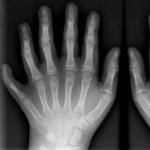 Polydactilia in newborn children
 Anti-cellulite cream: "For" and "against
 Causes of skin peeling on face
 What clothes and who can you wear it?
 How to reliably find out that the wife changes and whether this is needed by many wives change