The turbinates divide the lateral portion of the nasal cavity into three nasal passages: upper, middle and lower. Bony walls of the nasal cavity
The nasal cavity has paranasal sinuses, which communicate with various nasal passages (Fig. 50). So, in the upper nasal passage, the body cavity of the main bone and the posterior cells of the ethmoid bone open, in the middle nasal passage - the frontal and maxillary sinuses, the anterior and middle cells of the ethmoid bone. The lacrimal canal flows into the lower nasal passage.
Figure: fifty.
A - the outer wall of the nasal cavity with holes in the paranasal sinuses: 1 - the frontal sinus; 3 - opening of the frontal sinus; 3 - opening of the anterior ethmoid cells; 4 - hole maxillary sinus; 5 - holes of the posterior cells of the ethmoid bone; 6 - the main sinus and its opening; 7 - pharyngeal opening of the auditory tube; 8 - opening of the nasolacrimal duct. B - nasal septum: 1 - crista galli; 2 - lamina cribrosa; 3 - lamina perpendicularis ossis ethmoidalis; 4 - opener; 5 - hard palate; 5 - cartilago septi nasi.
Maxillary sinus (sinus maxillaris Highmori) located in the body upper jaw... It begins to be created from the 10th week of embryonic life and develops up to 12-13 years. In an adult, the volume of the cavity ranges from 4.2-30 cm 3, it depends on the thickness of its walls and less on its position. The shape of the sinus is irregular, it has four main walls. The anterior (in 1/3 of cases) or antero-external (in 2/3 of cases) wall is represented by a thin plate corresponding to the fossa canina. There is n on this wall. infraorbitalis together with the blood vessels of the same name.
The upper wall of the sinus is also the lower wall of the orbit. In the thickness of the wall there is canalis infraorbitalis, containing the mentioned neurovascular bundle. At the site of the latter, the bone may be thinned or have a gap. In the presence of a gap, the nerve and blood vessels are separated from the sinus only by the mucous membrane, which leads to inflammation of the infraorbital nerve in sinusitis. Usually, the top wall of the sinus is flush with the top of the middle nasal passage. N.N. Rezanov points to a rare variant when this wall of the sinus is low and the middle nasal passage is adjacent to the inner surface of the orbit. This is due to the possibility of penetration into the orbit of the needle during the puncture of the maxillary sinus through the nasal cavity. Often, the dome of the sinus extends into the thickness of the inner wall of the orbit, pushes the ethmoid sinuses up and back.
The lower wall of the maxillary sinus is represented by the alveolar process of the jaw, corresponding to the roots of the 2nd small and anterior large molars. The zone of the position of the roots of the teeth can protrude into the cavity in the form of an elevation. The bone plate separating the cavity from the root is often thinned, sometimes has a gap. These conditions favor the spread of infection from the affected tooth roots to the maxillary sinus, explaining cases of tooth penetration into the sinus at the time of its extirpation. The bottom of the sinus can be 1–2 mm above the bottom of the nasal cavity, at the level of this bottom or below it as a result of the development of the alveolar bay. The maxillary cavity rarely extends under the bottom of the nasal cavity, forming a small depression (buchta palatina) (Fig. 51).

Figure: 51. Paranasal sinuses, maxillary sinus.
A - sagittal cut: B - frontal cut; В - construction options - high and low position of the lower wall: 1 - canalis infraorbitalis; 2 - fissura orbitalis Inferior; 3 - fossa pterygopalatina; 4 - maxillary sinus; 5- ethmoid cells; 6 - eye socket; 7 - processus alveolaris; 8 - lower nasal concha; 9 - nasal cavity; 10 - buchta prelacrimalis; 11 - canalis infraorbitalis (deprived of the lower wall); 12 - buchta palatina; 13 - buchta alveolaris; G - frontal sinus on a sagittal cut; D - options for the structure of the frontal sinus.
The inner wall of the maxillary sinus is adjacent to the middle and lower nasal passages. The wall of the lower nasal passage is solid, but thin. Here it is relatively easy to puncture the maxillary sinus. The wall of the middle nasal passage has a webbed structure over a considerable extent and an opening that communicates the sinus with the nasal cavity. Hole length 3-19 mm, width 3-6 mm.
The posterior wall of the maxillary sinus is represented by the maxillary tubercle in contact with the pterygopalatine fossa, where n. infraorbitalis, ganglion sphenopalatinum, a. maxillaris with its branches. Through this wall you can approach the pterygopalatine fossa.
Frontal sinuses (sinus frontalis) are located in the thickness of the frontal bone, corresponding to the superciliary arches. They look like triangular pyramids with a downward base. Sinuses develop from 5-6 to 18-20 years. In adults, their volume reaches 8 cm 3. Upward, the sinus extends somewhat beyond the brow ridges, outward - to the outer third of the upper edge of the orbit or to the supraorbital notch and descends down into the nasal part of the bone. The anterior wall of the sinus is represented by the superciliary tubercle, the posterior wall is relatively thin and separates the sinus from the anterior cranial fossa, the lower wall forms part of the upper wall of the orbit and at the midline of the body - part of the nasal cavity, the inner wall is the septum separating the right and left sinuses. The top and side walls are absent, since its front and back walls converge at an acute angle. The cavity is absent in about 7% of cases. The partition separating the cavities from each other does not occupy a middle position in 51.2% (M.V. Miloslavsky). A cavity opens through a canal (canalis nasofrontalis) up to 5 mm in length into the middle nasal passage, in front of the opening of the maxillary sinus. In the frontal sinus, canalis nasofrontalis is formed at the bottom of its funnel. This promotes the drainage of mucus from the sinus. Tillo points out that the frontal sinus can sometimes open into the maxillary sinus.
Ethmoid sinuses (sinus ethmoidalis) are represented by cells corresponding to the level of the upper and middle turbinates, constituting the upper part of the lateral wall of the nasal cavity. These cells communicate with each other. On the outside, the cavities are delimited from the orbit by a very thin bone plate (lamina papyrocea). If this wall is damaged, air from the cells of the cavity can penetrate into the tissue of the periorbital space. The resulting emphysema gives rise to a protrusion of the eyeball - exophthalmos. From above, the cells of the sinus are delimited by a thin bony septum from the anterior cranial fossa. The anterior group of cells opens into the middle nasal passage, the posterior one - into the upper nasal passage.
Main sinus (sinus sphenoidalis) is located in the body of the main bone. It develops between the ages of 2 and 20. The sinus is divided into right and left by a septum along the midline. The sinus opens into the upper nasal passage. The hole lies 7 cm from the nostril along a line following through the middle of the middle turbinate. The position of the sinus made it possible to recommend that surgeons approach the pituitary gland through the nasal cavity and nasopharynx. The main sinus may or may not be present.
Lacrimal canal (canalis nasolacrimalis) is located in the zone of the lateral border of the nasal region (Fig. 52). It opens into the lower nasal passage. The opening of the canal is located under the anterior edge of the inferior turbinate on the outer wall of the nasal passage. It is 2.5-4 cm from the posterior edge of the nostril. The length of the lacrimal canal is 2.25-3.25 cm (N.I. Pirogov). The channel runs in the thickness of the outer wall of the nasal cavity. In the lower segment, it is limited by bone tissue only on the outside, on the other sides it is covered with the mucous membrane of the nasal cavity.

Figure: 52. Topography of the lacrimal passages.
1 - fornix sacci lacrimalis; 2 - ductus lacrimalis superior; 3 - papilla et punctum lacrimale superior; 5 - caruncula lacrimalis; 6 - ductus et ampula lacrimalis Inferior; 7 - saccus lacrimalis; 8 - m. orbicularis oculi; 9 - m. obliquus oculi inferior; 10 - sinus maxillaris; 11 - ductus nasolacrimalis.
A - cross section: 1 - lig. palpebrale medialis; 2 - pars lacrimalis m. orbicularis oculi; 3 - septum orbitale; 4 - f. lacrimalis; 5 - saccus lacrimalis; 6 - periosteum
Nasal cavity (cavum nasi)the septum divides into two identical halves, called the right and left halves of the nose. From the front, the nasal cavity communicates with the environment through the nostrils, and from the back through the choanae fromthe upper part of the pharynx - the nasopharynx.
Each half of the nasal cavity has four walls: medial, lateral, upper and lower. The nasal cavity begins with a vestibule, which, unlike its other parts, is lined with skin, which has a significant amount of hair, serving, to a certain extent, as a filter that retains large dust particles when breathing through the nose.
On the lateral wall of the nose (Fig. 4), three protrusions are clearly visible, located one above the other. These are the conchae nasales: lower, middle and upper (conchae nasalis inferior, media et superior). The base of the lower, largest turbinate is an independent bone, while the middle and upper turbinates are parts of the ethmoid bone.
A slit-like space is defined under each nasal concha - the nasal passage. Accordingly, there are lower, middle and upper nasal passages (meatus nasi inferior, medius et superior). The space between the free surface of the turbinates and the nasal septum forms a common nasal passage.
Figure: 4. Lateral wall of the nasal cavity.
1.Medium shell. 2. Joint of the maxillary sinus; 3.Frontal sinus; 4. Joint of the frontal sinus; 5. Lacrimal canal; 7. Lower nasal passage; 8. Middle nasal passage; 9.Upper turbinate; 10.Middle turbinate; 11.Inferior turbinate; 12. The mouth of the auditory tube; 13.Upper nasal passage; 14. Sphenoid sinus; 15.Justice of the sphenoid sinus; 16. Sieve plate; 17.Ophalous zone.
In addition to bone tissue in the submucosal layer of the turbinates, there is an accumulation of varicose venous plexuses (a kind of cavernous tissue), in which arterioles of small diameter flow into venules of a larger diameter. This makes it possible for the nasal conchas to increase in volume and narrow the lumen of the common nasal passage under the influence of certain stimuli, which contributes to a longer contact of the inhaled air with the mucous membrane filled with blood.
In the lower nasal passage under the anterior ends of the concha into the nasal cavity, the lacrimal canal opens, through which the tear flows. In the middle nasal passage, most of the paranasal sinuses open (maxillary, frontal, anterior and middle cells of the ethmoid labyrinth), therefore, sometimes the middle nasal passage is called the "mirror of the paranasal sinuses", since a purulent, catarrhal pathological process is manifested by characteristic secretions in the middle nasal passage ( fig. 5). On
the lateral wall of the middle nasal passage is a semilunar slit (hiatus semilunaris), which in the posterior part has an expansion in the form of a funnel (infundibulum ethmoidale). Into the lattice funnel anteriorly and upward

Fig. 5. Communication of the paranasal sinuses with the nasal cavity.
1.Inferior turbinate; 2. The opening of the lacrimal canal; 3. Lower nasal passage; 4. Middle turbinate. 5.Frontal sinus; 6. Joint of the frontal sinus; 7. Lattice bubble; 8.The joint of the maxillary sinus; 9.Upper turbinate; 10.Upper nasal passage; 11.Justice of the sphenoid sinus; 12. Sphenoid sinus; 13. Pharyngeal tonsil; 14. The pharyngeal opening of the auditory tube.
the excretory canal of the frontal sinus opens, and posteriorly and downward - the natural anastomosis of the maxillary sinus. In the middle nasal passage, the anterior cells of the ethmoid labyrinth open. The natural anastomosis of the maxillary sinus is covered by the uncinate process (processus uncinatus), so the anastomosis cannot be seen during rhinoscopy. In recent years, in connection with the introduction of endoscopic methods of rhinosurgery, it is necessary to know such details of the anatomical structure of the nasal cavity as the "ostiomeatal complex" - a system of anatomical structures in the region of the middle nasal passage (Fig. 6). It includes
. 
Fig. 6. Coronal section through the ostiomeatal complex.
1. Anastomosis of the frontal sinus; 2.Paper plate; 3.Middle turbinate; 4. Lattice bubble; 5. Middle nasal passage; 6. Funnel; 7. Hook-shaped process. 8.Joint of the maxillary sinus.
hook-shaped process, nasal roller cells (agger nasi), posteriorly - a large ethmoid vesicle (bulla ethmoidales) and the lateral surface of the middle nasal concha.
Medial wall the nasal cavity is represented by the nasal septum (septum nasi), consisting of two bone elements - the perpendicular plate of the ethmoid bone and the vomer, as well as the cartilaginous plate (quadrangular cartilage) and the part located in the vestibule of the nose, consisting of skin duplication - the movable part of the nasal septum (Fig. . 7).
The vomer is an independent bone in the shape of an irregular quadrangle. At the bottom, the opener adjoins the nasal crest of the palatine processes of the upper jaw and palatine bone. Its rear edge forms

Figure: 7. Septum of the nose.
1.Medial pedicle of the greater wing cartilage; 2. Quadrangular cartilage; 3. Nasal bone; 4. Frontal sinus; 5. Perpendicular plate of the ethmoid bone; 6. Sphenoid sinus. 7. Opener.
a septum between the right and left choans. The upper edge of the quadrangular cartilage forms the lower dorsum of the nose. This should be taken into account when surgery for curvature of the nasal septum - too high resection of the cartilage can lead to retraction of the nasal dorsum. IN childhood, as a rule, up to 5 years, the nasal septum is not curved, and in the future, due to the uneven growth of the bone and cartilaginous parts of the nasal septum, its deviation, expressed in varying degrees, occurs. In adults, more often in men, the curvature of the nasal septum is observed in 95% of cases.
Top wall the nasal cavity in the anterior sections is formed by the nasal bones, in the middle section - by the ethmoid plate of the ethmoid bone (lamina cribrosa). This is the narrowest section of the roof of the nasal cavity, only a few millimeters. This wall is very thin and with careless surgical interventions in the nasal cavity, damage to this thin plate can occur with the occurrence of nasal liquorrhea. With an associated infection, inflammation of the meninges (meningitis) is possible. The upper wall is pierced by a large number of small holes (about 25-30), passing into the nasal cavity fibers of the olfactory nerve, the anterior ethmoid nerve and the vein accompanying the ethmoid artery - a source of possible heavy nosebleeds.
Bottom wall the nasal cavity delimits the nasal cavity from the oral cavity, it is formed by the palatine process of the upper jaw and the horizontal plate of the palatine bone. The width of the bottom of the nasal cavity in an adult is 12-15mm, in a newborn - 7mm. Posteriorly, the nasal cavity communicates through the choanae with the nasal part of the pharynx. In a newborn, choanas have a triangular or rounded shape, measuring 6x6 mm, and by the age of 10 they double.
In young children, the nasal passages are narrowed by the turbinates. The inferior turbinate fits snugly to the bottom of the nasal cavity. Therefore, in young children, even a slight inflammation of the nasal mucosa leads to a complete shutdown of nasal breathing, a disorder of the sucking act.
The mucous membrane of the nasal cavity lines two conditionally divided zones - olfactory and respiratory. Along the entire length, the mucous membrane of the respiratory zone is firmly connected with the underlying bone and cartilage formations. Its thickness is about 1mm. The submucosal layer is absent. The mucous membrane of the nasal cavity contains ciliated epithelial cells, as well as a large number of goblet and basal cells. On the surface of each cell, there are 250 to 300 cilia, which perform from 160 to 250 vibrations per minute. These cilia oscillate in the direction of the posterior portions of the nasal cavity, towards the choanas (Fig. 8).

Fig. 8. Mucociliary transport scheme.
1.3 Mucus; 2.Cilia (cilia); 4.Microvilli.
In inflammatory processes, the cells of the ciliated epithelium can metaplase into the goblet and, like them, secrete nasal mucus. Basal cells contribute to the regeneration of the nasal mucosa. Normally, the nasal mucosa secretes about 500 ml of fluid during the day, which is necessary for the normal functioning of the nasal cavity. In inflammatory processes, the excretory capacity of the nasal mucosa increases many times. Under the cover of the mucous membrane of the nasal concha is a tissue consisting of a plexus of small and large blood vessels - a whole ball of dilated veins, resembling cavernous tissue. The walls of the veins are richly supplied with smooth muscles, which are innervated by the fibers of the trigeminal nerve and, under the influence of stimulation of its receptors, can contribute to the filling or emptying of the cavernous tissue, mainly of the inferior turbinates. Normally, both halves of the nose usually breathe unevenly during the day - either one or the other half of the nose breathes better, as if giving the other half a rest (Fig. 9).

Fig. 9. Nasal cycle on CT scan of the paranasal sinuses.
In the anterior part of the nasal septum, a special zone can be distinguished, with an area of \u200b\u200babout 1 cm 2, where the accumulation of arterial and especially venous vessels great. This bleeding area of \u200b\u200bthe nasal septum is called the locus Kiesselbachi, and it is from this area that nosebleeds most often occur (Fig. 10).

Figure: 10. Bleeding area of \u200b\u200bthe nasal septum.
1. Anterior and posterior ethmoid arteries. 2. Wedge-palatine artery; 3. Palatine artery; 4. Lip artery; 5.Kisselbach's place.
The olfactory region covers the upper parts of the middle shell, the entire upper shell and the upper part of the nasal septum located opposite it. Axons (non-fleshy nerve fibers) of olfactory cells in the form of 15-20 thin nerve filaments pass through the openings of the ethmoid plate into the cranial cavity and enter the olfactory bulb. Dendrites of the second neuron approach the nerve cells of the olfactory triangle and reach the subcortical centers. Further from these formations, the fibers of the third neuron begin, reaching the pyramidal neurons of the cerebral cortex - the central part of the olfactory analyzer.
Blood supply to the nasal cavity carried out from the maxillary artery, one of the terminal branches of the external carotid artery. The wedge-palatine (a. Sphenopalatina) departs from it, entering the nasal cavity through the hole of the same name approximately at the level of the posterior end of the middle shell. It gives branches for the lateral wall of the nose and the nasal septum, through the incisor canal, anastomoses with the great palatine artery and the artery of the upper lip. In addition, the anterior and posterior ethmoidal arteries (a. Ethmoidalis anterior et posterior) penetrate into the nasal cavity, extending from the ophthalmic artery, which is a branch of the internal carotid artery (Fig. 11).
Thus, the blood supply to the nasal cavity is carried out from the system of the internal and external carotid arteries, and therefore, the ligation of the external carotid artery does not always lead to stopping persistent nosebleeds.
The veins of the nasal cavity are located more superficially relative to the arteries and form several plexuses in the mucous membrane of the turbinates, the nasal septum, one of which - the Kisselbach place - was described earlier. In the posterior parts of the nasal septum, there is also an accumulation of venous vessels of a larger diameter. The outflow of venous blood from the nasal cavity goes in several directions. From the posterior parts of the nasal cavity, venous blood enters the pterygoid plexus (plexus pterigoideus), which in turn is associated with the cavernous sinus (sinus cavernosus), located in the middle cranial fossa. This can lead to the spread of the infectious process from the nasal cavity and nasal part of the pharynx into the cranial cavity.
From the anterior parts of the nasal cavity, venous blood flows into the veins of the upper lip, angular veins, which also through the superior orbital vein

Fig. 11. Blood supply to the nasal cavity.
1. Anterior ethmoid artery; 2. Posterior ethmoid artery; 3. Meningeal artery; 4. Wedge-palatine artery; 5.Maxillary artery. 6. Internal carotid artery .; 7.External carotid artery; 8. Common carotid artery; 9.Maxillary artery embolization site.
penetrate into the cavernous sinus. That is why with a boil located at the entrance to the nose, where there is hair, it is also possible for the infection to spread into the cranial cavity. Of great importance is the connection of the anterior and posterior veins of the ethmoid labyrinth with the veins of the orbit, which can cause the transition of the inflammatory process from the ethmoid labyrinth to the contents of the orbit. In addition, one of the branches of the anterior veins of the ethmoid labyrinth, passing through the ethmoid plate, penetrates into the anterior cranial fossa, anastomosing with the veins of the soft meninges... Due to the dense venous network with numerous anastomoses in the border areas, severe complications may develop, such as thrombophlebitis of the maxillofacial region, thrombosis of the veins of the orbit, thrombosis of the cavernous sinus, and the development of sepsis.
Lymphatic vesselsthey divert lymph to the posterior parts of the nasal cavity, penetrate into the nasal part of the pharynx, bypassing the pharyngeal openings of the auditory tubes from above and below, penetrate into the pharyngeal lymph nodes located between the prevertebral fascia and the fascia of the neck in loose tissue. Part lymphatic vessels from the nasal cavity are sent to deep cervical nodes. Suppuration of the lymph nodes during inflammatory processes in the nasal cavity, paranasal sinuses, and also in the middle ear can lead to the development of pharyngeal abscesses in childhood. Metastases in malignant neoplasms of the nasal cavity and ethmoid labyrinth also have a certain localization associated with the features of lymph outflow: an increase in lymph nodes along the internal jugular vein.
Innervation - in addition to the olfactory nerve (n.olphactorius), described earlier, the nasal mucosa is supplied with sensitive fibers I and II branches of the trigeminal nerve (n. Trigeminis). The peripheral branches of these nerves, innervating the region of the orbit and teeth, anastomose with each other. Therefore, there may be a radiating pain reaction from some zones innervated by the trigeminal nerve to others, for example from the nasal cavity to the teeth and vice versa.
Russian translation of the article "Illustrated Essay: Anatomical Variations of the Paranasal Sinuses in Computed Tomography. How Does It Help Surgeons in Endoscopic Surgery?"
The lateral wall of the nasal cavity contains protrusions, which are called the upper, middle and lower turbinates, they divide the nasal cavity into the upper, middle and lower nasal passages. The superior nasal passage is drained into the posterior ethmoidal cells, and the wedge-shaped sinuses are drained into it through the sphenoetmoidal pocket. The frontal sinuses are drained into the middle nasal passage through the frontal pockets and maxillary sinuses through the openings of the sinuses, as well as the anterior ethmoid cells through their openings. The nasolacrimal canal is drained into the lower nasal passage.
Ostiomeatal complex
Ostiomeatal complex (hereinafter referred to as OMK) includes the opening of the maxillary sinus, ethmoid funnel, anterior ethmoid cells, and the frontal pocket (Fig. 1A). These structures are called anterior sinuses. OMK is a key structure in the pathogenesis of chronic sinusitis. Ethmoid cells are key in the drainage of the anterior sinuses. They are susceptible to injury during surgery due to their close connection with the orbit and anterior skull base.
Nasal tubercle cell
Nasal tubercle cell - the most anterior ethmoid cell, which protrudes anteriorly into the lacrimal bone. It is located in front, lower in relation to the frontal pocket, and is bordered by the opening of the frontal sinus (Fig. 1B). A good examination of the frontal pocket is possible when the nasal tubercle cell is opened. Its size can directly affect the patency of the frontal pocket and the anterior parts of the middle nasal passage.
Frontal pocket
Frontal pocket is a narrow air-containing channel that communicates with the frontal sinus. The frontal pocket is a frequent site for all sorts of inflammatory processes. The walls of the canal are formed by the cells of the tubercle of the nose in front, the paper plate laterally, the middle turbinate medially (Fig. 1B). The pocket opens in 62% into the middle nasal passage, in 38% into the lattice funnel. On coronal scans, the pocket is defined above the nasal tubercle cell.
Lattice funnel
Lattice funnel bounded in front by the hook-shaped process, behind by the front wall of the ethmoid bulla, and by the lateral paper plate (Fig. 1A). It opens into the middle nasal passage medially through the lunate fissure. On coronal scans, the bulla is located above the lattice funnel. The mouth of the maxillary sinus opens at the bottom of the funnel.
The ethmoid fossa is a critical element of anatomy for two reasons. First, it is most sensitive to iatrogenic damage and, as a consequence, to the formation of CSF fistulas. Second, the anterior ethmoid artery is at risk of injury, which can lead to uncontrolled bleeding into the orbit. In endoscopic surgery, intracranial injury can occur on the side where the ethmoid fossa is located below (Fig. 2).
The depth of the olfactory pit is determined by the height of the lateral lamella of the sieve plate, which is part of the ethmoid bone. In 1962, Keros classified the depth of the olfactory pit into three types: Keros 1, when the pit is less than 3 mm deep (Fig. 3), Keros 2, when the pit is 4-7 mm deep (Fig. 4), Keros 3, when the pit is 8 -16 mm deep (Fig. 5). Keros type 3 is the most dangerous for iatrogenic damage.
Onodi cells
Onodi cells are the posterior ethmoidal cells that protrude into the wedge-shaped sinuses (Fig. 6) and may even reach the optic nerve. When Onodi cells adjoin or surround the optic nerve, the nerve is at risk if these cells are surgically removed. This results in incomplete sphenoidectomy.
According to radiopedia.org, Onodi cells are sphenoetmoidal air cells, which are also defined as the most posterior ethmoidal cells that protrude posteriorly, upward and lateral to the sphenoid sinuses, located in close proximity to the optic nerve and the internal carotid artery. They often extend to the anterior inclined processes; it is important that the airiness of the anterior inclined process may be due simply to such a variant of the anatomy of the wedge-shaped sinus and does not necessarily indicate the presence of an Onodi cell.
Sphenoid sinus septum is attached to the wall containing the protrusion of the internal carotid artery, so arterial damage may be caused by removal of this sinus septum (Fig. 7). The artery can prolapse into the sinus in 65-72% of cases. There may be dehiscence or absence of the bone wall between the artery and sinus in 4-8% of cases.
Sinus agenesis can also be seen (Fig. 8).
The pterygoid canal (Fig. 9) or the maxillary nerve sulcus (Fig. 10) can prolapse into the sphenoid sinus, contributing to trigeminal neuralgia due to sinusitis.
Pneumatization of the anterior tilted processes (Fig. 9) is associated with type 2 and type 3 optic nerve positions and predisposes to nerve damage during endoscopic surgery.
Variants of the relationship between the optic nerve and the posterior paranasal sinuses
Optic nerve carotid arteries and the Vidian canal are formed before the appearance of the paranasal sinuses and contribute to congenital variants of the structure of the walls of the wedge-shaped sinuses. Delano, et al. divide the relationship between the optic nerve and the posterior paranasal sinuses into 4 groups:
- Type 1: The most common type, found in 76% of cases. In this case, the optic nerves adjoin the sphenoid sinus without the formation of depressions in its walls or contact with the posterior ethmoid cells (Fig. 11).
- Type 2: The optic nerves are adjacent to the sphenoid sinus, and the walls of the sinus deepen without contact with the posterior ethmoid cells (Fig. 12).
- Type 3: the nerves pass through the wedge-shaped sinuses, with at least half the circumference of the nerve being surrounded by air (Fig. 13)
- Type 4: nerves adjacent to the wedge-shaped sinus and posterior ethmoidal cells (Figs. 14 and 15).
Delano, et al. found that in 85% of cases pneumatized anterior tilted processes were associated with type 2 or 3 optic nerve position, while 77% showed dehiscence of the nerve canal wall (Fig. 16), which is associated with an increased risk of optic nerve injury with endoscopic surgery.
The sphenoid sinus septa may attach to the optic canal wall, predisposing to nerve injury during surgery (Fig. 17).
Middle turbinate options
The normal curvature of the middle turbinate is medial. When the bend is lateral, this is called the paradoxical bend of the middle turbinate (Fig. 18). Most authors agree that a paradoxically curved middle turbinate may be a contributing factor to sinusitis.
Сoncha bullosa is an aerated concha, usually the middle turbinate. When pneumatization involves the bulb of the middle turbinate, this condition is called concha bullosa (Fig. 19). If pneumatization involves the attachment of the middle turbinate to the base of the skull, this condition is called lamellar concha (Fig. 20).
Hook-shaped process options
On coronal scans, it can be determined that the posterior section of the uncinate process is attached to the inferior turbinate at the bottom, while the posterior edge of the process remains free. The anterior section of the uncinate process is attached to the base of the skull from above, to the middle turbinate medially, to the paper plate or to the cusp of the nasal tubercle laterally.
The uncinate process can be medialized, lateralized, pneumatized, or curved. Medialization occurs as a large ethmoid bulla is present. Lateralization occurs when trellised funnel obstruction occurs. Pneumatization of the uncinate process (processus bulla) (Fig. 21) occurs in 4% of the population and rarely leads to obstruction of the ethmoid funnel.
Haller's cells
Haller's cells, they are infraorbial ethmoid cells (Fig. 22), located along the medial wall of the maxillary sinus and the lowest portion of the paper plate, below the ethmoid bulla, lateral to the uncinate process. These cells can narrow the ethmoid funnel and the mouth of the maxillary sinus, contribute to the appearance of recurrent maxillary sinusitis.
According to radiopedia.org, Haller's cells (infraorbital ethmoidal cells or maxilloetmoidal cells) are extramural ethmoidal cells that project towards the inferior medial edge of the orbit and are present in about 20% of patients (2-45%). Their importance increases when they are affected by the inflammatory process, the inflammation from them can move into orbit; cells can narrow the ethmoid funnel or the mouth of the maxillary sinus, if the cells are large, and contribute to sinus obstruction during inflammation; when resecting a Haller's cell, the orbit may be damaged.
Lattice bull
The largest and most prominent front trellis cage is called lattice bull... It is located lateral to the paper plate. The bulla can merge with the base of the skull from above and the basal plate of the middle turbinate in the back. On coronal scans, it is located upward from the lattice funnel (Fig. 23). The decrease in the degree of pneumatization of the bulla varies, and the absence of pneumatization of the bulla is referred to as torus ethmoidalis. The giant bulla can fill the middle nasal passage and is located between the uncinate process and the middle turbinate.
Air cells of the posterior-superior portion of the nasal septum
Air cells can be located in the back-upper portion of the nasal septum and connect with the wedge-shaped sinus (Fig. 24). Inflammatory processes that occur in the paranasal sinuses can also affect these cells. These cells can resemble a cephalocele.
Cockscomb
Cockscomb can be pneumatized, and the crest can communicate with the frontal pocket, cause obstruction of the frontal sinus opening and lead to chronic sinusitis or mucocele formation. It is important to detect and distinguish this variant of the ethmoid cell before surgery in order to avoid penetration into the anterior cranial fossa.
In the outer nose distinguish between the nose bridge, passing into the back of the nose, formed by the convergence of its lateral surfaces (lateral slopes). The bridge of the nose ends with the tip of the nose. The lower part of the lateral surfaces is formed by the wings of the nose, which are separated from the lateral surface of the wing, and from the upper lip-nasolabial groove. The nasal openings, nostrils (nares), are separated by the movable part of the nasal septum.
Bone skeleton of the external nose consists of the nasal bones and the frontal processes of the upper jaw. The upper ends of the nasal bones at the junction with the nasal processes of the frontal bone form the root of the nose (nose bridge). The lateral edges of the nasal bones are connected along the entire length with the frontal processes of the upper jaw, forming the lateral surface of the nose, while the inner edges are connected to each other, and below with triangular cartilage; the frontal processes of the upper jaw are connected at the top through a suture with the frontal bone, medially with the nasal bones, and laterally form part of the inner and lower edges of the orbit.
Nasal bones, the frontal processes of the upper jaw and the anterior lower nasal spine of the upper jaw limit the pear-shaped opening on the macerated skull, which is naturally closed by the cartilaginous framework of the nose. The latter consists of unpaired quadrangular cartilage adjacent to the anterior-lower edge of the bony nasal septum, and paired lateral (triangular) and large and small wing cartilages. There are many sebaceous glands at the end of the outer nose. Curving over the edge of the nasal openings, the skin extends to the vestibule of the nasal cavity, where it is supplied with hairs.
Vessels of the external nose represented by the branches of the external jaw artery and the orbital artery, anastomosed with each other. All arteries are directed to the coccyx of the nose, which is rich in blood supply. The veins of the external nose anastomose with the veins of the nasal cavity and flow into the anterior facial vein. Insertion of the muscles of the external nose is carried out by branches of the facial nerve, and the skin is supplied by its first and second branches of the trigeminal nerve.
Lateral nasal wall the most complex in its structure. It is formed (going from front to back) by the inner surface of the nasal bone, the inner surface of the frontal process, to which the lacrimal bone adjoins above and behind, and the medial (nasal) surface of the upper jaw body, on which there is a large round or oval opening (hiatus maxillaris) leading into the maxillary sinus.
Further into the composition of the wall the vertical plate of the palatine bone enters, limiting the posterior inferior edge of the sinus opening, and, finally, behind the lateral wall is closed by the medial plate of the main bone. Between the processes of the upper end of the vertical plate of the palatine bone and the body of the main bone there is an opening - foramen sphenopalatinum, connecting the nasal cavity with the pterygopalatine fossa.
The anatomy of the nose and paranasal sinuses is of great clinical importance, since not only the brain is located in the immediate vicinity of them, but also many great vessels, which contribute to the rapid spread of pathogenic processes.
It is important to understand exactly how the structures of the nose communicate with each other and with the surrounding space in order to understand the mechanism of development of inflammatory and infectious processes and to prevent them qualitatively.
The nose, as an anatomical formation, includes several structures:
- external nose;
- nasal cavity;
- paranasal sinuses.
External nose
This anatomical structure is an irregular pyramid with three faces. The outer nose is very individual in appearance and has a wide variety of shapes and sizes in nature.
The backrest delimits the nose from the upper side, it ends between the eyebrows. The top of the nasal pyramid is the tip. The lateral surfaces are called wings and are clearly separated from the rest of the face by nasolabial folds. Thanks to the wings and the nasal septum, a clinical structure such as the nasal passages or nostrils is formed.
External nose structure
The outer nose includes three parts
Bone skeleton
Its formation occurs due to the participation of the frontal and two nasal bones. The nasal bones are bounded on both sides by processes extending from the upper jaw. Bottom part of the nasal bones participates in the formation of the pear-shaped opening, which is necessary for the attachment of the external nose.
Cartilaginous part
Lateral cartilage is required for the formation of the lateral nasal walls. If you go from top to bottom, then the adjoining of the lateral cartilages to the large cartilages is noted. The variability of small cartilages is very high, since they are located near the nasolabial fold and can differ from person to person in number and shape.
The septum of the nose is formed by the quadrangular cartilage. The clinical significance of cartilage is not only in hiding the inner part of the nose, that is, in organizing a cosmetic effect, but also in the fact that, due to changes in the quadrangular cartilage, a diagnosis of curvature of the nasal septum may appear.

Soft tissues of the nose
The person does not have a strong need for the muscles surrounding the nose to function. Basically, muscles of this type perform mimic functions, helping the process of identifying smells or expressing an emotional state.
The skin adheres strongly to the surrounding tissues, and also contains many different functional elements: glands that secrete fat, sweat, hair follicles.
Hair blocking the entrance to the nasal cavities performs a hygienic function, being additional filters for air. Due to hair growth, the formation of the nasal threshold occurs.
After the threshold of the nose, there is a formation called the intermediate girdle. It is tightly connected with the perchondral part of the nasal septum, and when it deepens into the nasal cavity, it transforms into a mucous membrane.
To correct a deviated nasal septum, an incision is made exactly in the place where the intermediate girdle is tightly connected to the perchondral part.

Circulation
The facial and orbital arteries provide blood flow in the nose. Veins follow the course of arterial vessels and are represented by external and nasal veins. The veins of the nasolabial region merge in anastomosis with the veins that provide blood flow in the cranial cavity. This is due to the angular veins.
Because of this anastomosis, easy penetration of infection from the nasal region into the cranial cavity is possible.
Lymph flow is provided through the nasal lymphatic vessels, which flow into the facial, and those, in turn, into the submandibular.
The anterior ethmoid and infraorbital nerves provide sensitivity to the nose, while the facial nerve is responsible for muscle movement.
The nasal cavity is limited to three formations. It:
- anterior third of the cranial base;
- eye sockets;
- oral cavity.
The nostrils and nasal passages in front are the limitation of the nasal cavity, and posteriorly it passes into the upper part of the pharynx. The places of passage are called khoans. The nasal cavity is divided by a nasal septum into two approximately identical components. Most often, the nasal septum can deviate slightly to either side, but these changes do not matter.

The structure of the nasal cavity
Each of the two components has 4 walls.
Inner wall
It is created through the participation of the nasal septum and is divided into two sections. The ethmoid bone, or rather its plate, forms the posterior-upper section, and the vomer, the posterior-inferior section.
Outer wall
One of the complex formations. Consists of the nasal bone, the medial surface of the bone of the upper jaw and its frontal process, the lacrimal bone adjacent to the back, as well as the ethmoid bone. The main space of the posterior part of this wall is formed due to the participation of the bone of the palate and the main bone (mainly, the inner plate belonging to the pterygoid process).
The bony part of the outer wall serves as a place for the attachment of three turbinates. The bottom, vault and shells participate in the formation of a space called the common nasal passage. Thanks to the turbinates, three nasal passages are also formed - the upper, middle and lower.
The nasopharyngeal passage is the end of the nasal cavity.
Upper and middle concha

Nasal conch
Formed due to the participation of the ethmoid bone. The outgrowths of this bone also form the vesicle shell.
The clinical significance of this shell is due to the fact that its large size can interfere with the normal process of breathing through the nose. Naturally, breathing becomes difficult from the side where the vesicle is too large. Its infection must also be taken into account in the development of inflammation in the cells of the ethmoid bone.
Bottom sink
It is an independent bone that is attached to the crest of the maxillary bone and the bone of the palate.
The lower nasal passage has in its anterior third the orifice of a canal intended for the outflow of tear fluid.
Nasal conchas are covered soft tissueswhich are very sensitive not only to the atmosphere, but also to inflammation.
The middle passage of the nose has passages to most of the paranasal sinuses. An exception is the main sinus. There is also a semilunar slit, the function of which is to provide communication between the middle course and the maxillary sinus.
Top wall
The perforated plate of the ethmoid bone provides the formation of the nasal vault. The holes in the plate give the olfactory nerves a passage into the cavity.
Bottom wall

Blood supply to the nose
The bottom is formed due to the participation of the processes of the maxillary bone and the horizontal process of the bone of the palate.
The nasal cavity is supplied with blood by the main palatine artery. This same artery gives several branches for the blood supply to the wall located behind. The anterior ethmoid artery supplies blood to the lateral nasal wall. The veins of the nasal cavity merge with the facial and ocular veins. The eye branch has branches leading to the brain, which is important in the development of infections.
The deep and superficial network of lymphatic vessels ensures the outflow of lymph from the cavity. The vessels here communicate well with the spaces of the brain, which is important for accounting for infectious diseases and the spread of inflammation.
The mucous membrane is innervated by the second and third branches of the trigeminal nerve.
Paranasal sinuses
The clinical significance and functional properties of the paranasal sinuses are enormous. They work in close contact with the nasal cavity. If the sinuses are exposed to an infectious disease or inflammation, this leads to complications on important organs located in the immediate vicinity.
The sinuses are literally littered with a variety of holes and passages, the presence of which contributes to the rapid development of pathogenic factors and the aggravation of the situation in diseases.

Paranasal sinuses
Each sinus can cause infection to spread into the cranial cavity, eye damage, and other complications.
Maxillary sinus
Has a pair, located deep in the bone of the upper jaw. Sizes vary greatly, but the average is 10-12 cm.
The wall within the sinus is the lateral wall of the nasal cavity. The sinus has an entrance to the cavity located in the last part of the lunate fossa. This wall is endowed with a relatively small thickness, and therefore it is often pierced in order to clarify the diagnosis or conduct therapy.
The wall of the upper part of the sinus has the smallest thickness. The posterior sections of this wall may not have a bone base at all, making do with cartilaginous tissue and many clefts of the bone tissue. The thickness of this wall is penetrated by the inferior orbital nerve canal. The infraorbital foramen opens this canal.
The channel does not always exist, but this does not play any role, since if it is absent, then the nerve passes through the sinus mucosa. The clinical significance of such a structure is that the risk of developing complications inside the skull or inside the orbit increases if a pathogenic factor affects this sinus.
From below, the wall represents the holes of the most posterior teeth. Most often, the roots of the tooth are separated from the sinus by only a small layer of soft tissue, which is a common cause of inflammation if the condition of the teeth is not monitored.
Frontal sinus
It has a pair, located deep in the forehead bone, in the center between the scales and the plates of the part of the orbits. The sinuses can be delimited with a thin bone plate, and not always equally. Displacement of the plate to one side is possible. Holes may exist in the plate, providing communication between the two sinuses.
The sizes of these sinuses are variable - they may be absent altogether, or they can be hugely distributed throughout the frontal scales and the base of the skull.
The wall in front is the exit site for the nerve of the eye. The exit is provided by the presence of a notch above the eye socket. The notch cuts the entire upper part of the eye orbit. In this place, it is customary to open the sinus and trepanopuncture.

Frontal sinuses
The bottom wall is the smallest in thickness, which is why the infection can quickly spread from the sinus to the eye orbit.
The wall of the brain provides separation of the brain itself, namely the lobes of the forehead from the sinuses. It is also the site of infection.
A canal running in the frontal-nasal region provides an interface between the frontal sinus and the nasal cavity. The anterior cells of the ethmoid labyrinth, which have close contact with this sinus, often intercept inflammation or infection through it. Also, through this connection, tumor processes spread in both directions.
Lattice maze
It is cells separated by thin partitions. The average number of them is 6-8, but it can be more or less. The cells are located in the ethmoid bone, which is symmetrical and unpaired.
The clinical significance of the ethmoidal labyrinth is due to its close location to important organs. Also, the labyrinth can be adjacent to the deep parts that form the skeleton of the face. The cells located at the back of the labyrinth are in close contact with the canal in which the optic nerve runs. Clinical diversity seems to be an option when the cells serve as a direct pathway for the canal.
Diseases affecting the labyrinth are accompanied by a variety of pains that differ in localization and intensity. This is due to the peculiarities of the innervation of the labyrinth, which is provided by the branch of the orbital nerve, called the nasal nerve. The ethmoid plate also provides a path for the nerves needed for the sense of smell to function. That is why, if there is swelling or inflammation in this area, olfactory disturbances are possible.

Lattice maze
Main sinus
The sphenoid bone with its body provides the location of this sinus just behind the ethmoid labyrinth. The choanae and the vault of the nasopharynx will be located on top.
In this sinus there is a septum, which has a sagittal (vertical, dividing the object into the right and left parts) location. She, most often, divides the sinus into two unequal lobes and prevents them from communicating with each other.
The wall in front is a pair of formations: ethmoid and nasal. The first falls on the area of \u200b\u200bthe labyrinth cells located posteriorly. The wall is characterized by a very small thickness and, thanks to a smooth transition, almost merges with the wall from below. In both parts of the sinus there are small rounded passages that make it possible for the sphenoid sinus to communicate with the nasopharynx.
The back wall has a frontal position. The larger the size of the sinus, the thinner this septum, which increases the likelihood of injury during surgical interventions in this region.
 The wall from above is the bottom area of \u200b\u200bthe sella turcica, which is the location of the pituitary gland and the nerve cross that provides vision. Often if inflammatory process affects the main sinus, it spreads to the intersection of the optic nerve.
The wall below is the vault of the nasopharynx.
The walls on the sides of the sinus are closely adjacent to the bundles of nerves and blood vessels that are located on the side of the sella turcica.
In general, infection of the main sinus can be called one of the most dangerous. The sinus is closely adjacent to many structures of the brain, for example, with the pituitary gland, subarachnoid and arachnoid membranes, which simplifies the spread of the process to the brain and can be fatal.
Pterygopalatine fossa
Located behind the tubercle of the mandibular bone. A large number of nerve fibers pass through it, therefore the significance of this fossa in the clinical sense is difficult to exaggerate. A large number of neurological symptoms are associated with inflammation of the nerves passing through this fossa.
It turns out that the nose and the formations that are closely related to it are a completely complex anatomical structure. The treatment of diseases affecting the nasal system requires maximum care and caution from the doctor due to the close location of the brain. The main task of the patient is not to start the disease, bringing it to a dangerous border, and to seek medical help in a timely manner.
We also recommend
 State and municipal debt
 Which section of the balance sheet reflects debts to the budget?
 Cancellation of reporting for individual entrepreneurs
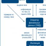 Accounting for cash transactions in brief Which account is intended for accounting of cash transactions
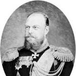 Who canceled the poll tax in Russia?
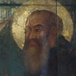 Ivan the Terrible - biography, information, personal life