Gigro (cyst synovial bag, synovial cyst, tendon gangliy). Hygroma (synovial cyst, gangliy) wrists, brushes, hands, feet, knee and other
The hygroma is a benign cystic formation in the form of a capsule filled with a transparent jelly-shaped mass. The clinically hygroma can be attributed to a rounded tumor, which is covered with normal skin on top. Gigrome size ranges from 5 to 30 cm in diameter. Sometimes there are both large sizes.
Causes of hygromy
To date, the cause of the occurrence of hygromas is not fully found out. It is often possible to observe a close relationship between the neoplascence and the recently obtained severe physical injuries, however, the disease is often developing at all without visible causes.
In some cases, cystic education is a consequence of chronic inflammatory process In the synovial bags that surround the joint (bursitis) or the tendon attached to the joint (tendovaginitis).
Preferably, the hygroma is developing in places that are subject to constant traumatization or pressure (poorly selected shoes, professional activities, etc.). The mechanical pressure is most susceptible to synovial bags located superficially, that is, on the back of the foot and the back surface of the ray-tank joint, therefore the risk of developing hygromas, foots or hygromas, but it happens that cyst is formed in other places.
According to statistics, men are more susceptible to the disease. It happens that the hygroma flows into the articular cavity and becomes visually invisible. But after a while, as a rule, it appears again.
On the human body, the hygroma may be present for quite a long time without causing any discomfort. It happens that a person lives all his life with this cystic education, not paying almost no attention to him. If the disease begins to cause certain inconveniences, causes pain when moving, is actively incremented in size or has a non-source species, the issue of operational removal of hygromas should be resolved.
Types of hygroma
The most common types of disease is a hyigrome of a brush, a hyigrome of the foot and a higer of the joint.
The hyigrome of the brush is formed, as a rule, in a plug--phalange articular area, on the blow muscles of the fingers or in the area of \u200b\u200bthe beam-up joint.
Higher Sustav is preferably painless cystic education. However, the hygroma joint often interferes with the patient to freely perform flexor-extensive movements, lead the familiar lifestyle, has to limit the daily burden on the joints.
The hyigrome of the foot is preferably localized in the field of ankle or on the back of a plus-phalange bone. Over time, the hygroma increases and compacted, which prevents the person to wear shoes and walk normally. In addition, if the hygroma is in a plus-phalangeal bone, it must be in a uniform state, otherwise the development of complications is possible.
Changes in tissues occurring in the hygroma
Most often, the hygroma looks like a watering bag of a synovial bag with a slightly pronounced inflammatory process. Sometimes blood is mixed with fluid inside the hygromas. As a rule, the walls of formation are thickened and soldered by surrounding tissues, often have a cartilage density.
With an active inflammatory process, the inner surface of the formation is covered by cellular growths, which leads to the formation of jumpers, heavy and pockets, which share the hygroma area into separate cameras. Small hygromas are usually filled with thick mass, and large-sulfuric fluid with cholesterol cholesterol and crystals.

Symptoms of hygromes
With small hygromy sizes, the patient, as a rule, does not complain about inconvenience. As education increases in size, the hygroma may cause pain that is significantly enhanced by physical Loads. If the tumor squeezes the blood vessels and nerves, there may be disturbances of sensitivity, blood stagnation in veins and pain in the course of nerves.
If the inflammatory process does not occur in the formation, then when palpation is a hygroma, it looks like a soft-elastic, shifted during tackling, rounded and slightly painful education.
In the event of an opening of the cyst as a result of injury or spontaneous, its contents begged through the resulting hole. If an infectious process occurs, it is accompanied by redness and swelling. Despite the fact that the hygroma does not threaten the lives of patients, they turn to the doctor about the treatment of hygromes, as the formation causes rather unpleasant painful sensations.
Diagnosis of hygromes
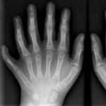
If the hygroma is located in superficial tissues, then its diagnosis does not represent special difficulties. In some cases, a differentiated diagnosis with an abscess, anneurysm of artery, some benign and malignant tumors can be prescribed. To completely eliminate these diseases, it is necessary to make a radiographic study.
Hygroma treatment
On the initial stage Diseases Treatment of hygromes, as a rule, is reduced to the use of mud and paraffin applications, physiotherapeutic and thermal procedures (electrophoresis with iodine, ultraviolet irradiation, etc.). Pressure (punctures) of the hygroma, with subsequent suction of the liquid and the introduction of glucocorticoid hormones in the resulting cavity of glucocorticoid hormones in the resulting cavity. After that, the affected area is tightly bandaged. However, such treatment of hygromas is effective only when a person can be released from work related to continuous damage to the synovial bag for a long period of time. Otherwise, in most cases, the disease can return again, as, despite the fact that the cavity falls, the sheath of the hygroma remains at the same place.
If the hygroma has suppuration, conduct its puncture, followed by peeks and administration to the resulting cavity of antibiotics. If necessary, the tumor can be opened, and the synovial bag is squeezed with a sharp spoon, after which they drain the wound.
If conservative treatment does not bring a strong result, the hygroma still has an inesthetical species and causes pain in the patient, continues to grow, the removal of hygromas is shown.
Bursectomy, or hygroma removal, is a complete excision of a synovial bag. After removing the hygromas on the affected area, 2-3 weeks are placed in order to immobilize the joint for the time of formation of the scar.
Prevention hygroma
In order to prevent the occurrence of a hygromy, it is necessary to avoid constant trauma of certain parts of the body during everyday life And work, wearing comfortable clothes and shoes. It is also recommended to conduct treatment of diseases that contribute to the formation of a hygromy.
With long-term exposure to the same area in the tissues, a benign cystic education appears - hygroma.
Pathology, most often, arises on the palms or in the field of ray-taking joint, because of which it is considered "professional" peculiar to pianists and labels.
So, what kind of disease is a hygroma, what are the reasons for its appearance and how to get rid of this neoplasm?
Description
A benign cyst consists of a dense wall, inside which a viscous jelly-like serous fluid with mucus impurities.
With the same probability, an adult and young man may appear, but young women are most susceptible to him. Located a tumor in the joint area.
About 50% of benign cystic formations in the field of leaky joints fall on hygromas.
Despite the fact that the disease is amenable to treatment, exists high risk Recurious.
The hygroma (code on the ICD 10 - M71.3) has the following locations:
- the surface of the ray-tank joint;
- ankle joint;
- knee-joint;
- elbow fold;
- the phalange surface of the hands (occurs in people whose profession implies a high load on the hands).
Types of pathology
There are two types:
- single-chamber - an inelastic egg, inside of which a liquid with mucin;
- the multi-chamber is elastic, with side branches, can expand inside the tissues.
Causes of hygromy
At the moment there is not a single full-fledged theory of the causal relationship of the prerequisites and the development of this neoplasm (none of them can describe all the occurrence).
The only reason for the occurrence of practical experience is - dangerous tendovaginitesPeople characteristic of people whose professional activities include multiple repetition of the same type of movements (pianist, packing, programmer, and so on).
Factors contributing to the development of the disease:
- heredity (blood relatives of a patient suffering from a hygroma fall into a risk group);
- primary and repeated injuries of the joints;
- activities associated with regular repetition of the same type of movements (professional musicians, programmers and other people working at the computer).
Location Classification
| Location | Symptoms and signs |
| Head | As a result of the crank-brain injuries, cerebrospinal fluid accumulates, which puts pressure on the brain. Subdural education is acute, subacute and chronic. Chronic is considered a hygroma, a period of 2 weeks. |
| Wrist, back of the brush | The most common type of pathology, causes strong inconvenience, as it prevents labor and household activities. The patient is hampered by flexion and extension of the joint, pain occurs. Required for treatment surgical intervention. |
| White Susta | It is a fibrin capsule. The neoplasm has a circle form (diameter up to 6 cm), located under the skin. On the initial stage The cyst does not hurt, but with growth begins to put pressure on the vessels, making it difficult. With severe pain syndrome, an operation is necessary. |
| Foot | Appears on the outside of the phalange bones or the ankle. Education prevents walking, carrying shoes. The greatest danger representing the disease is to transmit the nerve endings in the field of lesion. |
| Knee-joint | The cyst is formed on the foot as a result of the accumulation of fluid in the cavity of the synovial bag. The most susceptible to the disease people experiencing high loads on their feet (athletes, sellers, couriers). |
| Tendon | Education is developing from the articular synovial shell and tendon tissue. The hygroma causes significant discomfort, especially if an injury appeared. Pain syndrome Enhanced when driving or mechanical exposure. |
Symptomatics of the disease
Depending on the size of the neoplasm, clinical manifestations may be different. Small hygromas are practically invisible for patients and cause a minimum of discomfort.
Usually cyst does not exceed 2-5 cm in diameter. If the tumor is increased, the synovial bag is stretched, causing pain and inconvenience.

When squeezing the vessels and nerve trunks, symptoms appear:
- tingling the skin at the place of lesion, numb;
- neurological pains;
- stagnation on the affected area.
What a hygrome looks like: a cystow-shaped tumor has a spherical shape. To the touch formation is dense and elastic due to the jelly-like content.
The hyigrome in the photo will be clearly visible, since the neoplasm is emphasized in the joint area.
At the launched stages, the disease causes a violation of venous blood circulation on an affected area arising from the squeezing of large veins. Manifests itself as follows:
- the appearance of edema is below the hygroma;
- "Syushia" of the skin;
- pain syndrome;
- limiting the mobility of the joint.
In addition to vessels, nervous trunks are squeezed, which also leads to significant discomfort and pain.
In 35% of cases, the disease may not show symptoms. This happens if the formation is located under the ligament or for a long time does not increase in size, and is detected only when pain appears.
Possible complications
Complications are possible when opening hygromas (spontaneous, traumatic). In this case, for a long time, the contents of the cyst appears through the hole that appears.
Damage to education fraught with inflammatory reaction, and when infections getting nudenia.

The clinical picture is characterized by signs of the inflammatory process:
- local temperature increase;
- redness of the skin;
- painful syndrome;
- swimming the damaged area.
Diagnosis of hygromes
To make a diagnosis, the doctor requires a collection of information about the disturbing symptoms and inspection of the patient.
Since the signs of the disease are typical, it can be confused with other similar pathologies. Treatment of hygromes is carried out by a traumatologist.
Diagnosis of the disease In order to differentiate it with other pathologies, the following medical examinations are carried out:
- x-ray;
- ultrasound examination (ultrasound);
- puncture with biopsy;
If, as a result of the study, other diseases were excluded and the growth of bacteria in the bioptate did not occur (that is, the contents of the cyst sterile), the doctor puts the diagnosis of the hygromy. The final conclusion on the patient's condition a specialist is based on the study of an X-ray.
Treatment
To understand how to treat a hygroma, you need to proceed from education size. Small tumors are well amenable to conservative therapy (treatment without surgery). Large cyst discomfort is made to remove surgically.
Conservative methods
In pathologies of a small size, a patient may assign a visit to a physiotherapy office. Through warming procedures and electrophoresis, symptoms are eliminated and the tumor size decreases. The disease requires the binting of the affected area, reduced motor activity.
As practice shows, the procedures are ineffective in the treatment of hygromas, but allow you to restrain its development and reduce discomfort. With such a treatment, there is a risk of recurrence.
Also, physiotherapeutic procedures are prescribed to restore after invasive treatment, pumping fluid from the neoplasm.

Professional massage contributes to an outflow of a synovial fluid in the direction of the articular slit, which leads to a decrease in the tumor in size and improving the patient's well-being.
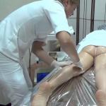
Effective treatment method is puncture. By puncture, the contents of the cyst are removed, and the sclerosing substance is introduced into the resulting cavity. It glues the walls of the capsule, thereby prevents the re-accumulation of fluid. Upon completion of the procedure, a pressure bandage is superimposed on the point of puncture, which ensures reliable gluing walls. The joint is covered with plaster to reduce the production of fluid and immobilize it.
Surgery
Indications for operational removal of hygromes:
- pain syndrome, which manifests itself both when driving and at rest;
- the impossibility of full motor activity;
- accelerated cyst growth.
Operation can be carried out for cosmetic purposes.
In the postoperation period, it is necessary to ensure peace of the body where there was a neoplasm. This will help reduce the recovery period.

With a favorable scenario, rehabilitation takes no more than one week, but the exact amount of time required to return to normal operation is determined individually. It all depends on the volume of operation, the availability of complications.
The swelling of the operated region, the presentation of Sukrovitsy or Mouth through the seams may indicate the teething of the skin with suture materials, the discrepancy of the wound. In this case, a re-operation is required.
In the course of surgical intervention, dead tissues will be removed, the focus of inflammation is eliminated. Rehabilitation will be required to two weeks.
Folk treatments
Many patients prefer to get rid of hygromy by folk remedies. The most effective of them is the use of compresses and decoctions based on plant components.

To eliminate small neoplasms, you can use a copper coin:
- it must be attached to the affected place;
- tightly tied, without sneaking fabrics.
After a few days, the hygroma is absorbed.
Compresses and rubbing
How to remove a hygroma at home? For this you can use folk treatments. However, it is important to comply with the methodology and regularly perform the necessary operations in applying methods of alternative medicine.
Recipe 1: Rubbing from Physalis
- on the meat grinder to crush the fruit of Physalis;
- at night, apply the composition to the affected area;
- covered with cotton cloth, cover with cellophane;
- fix the bandage;
- in the morning wash the compress;
- in the evening, wash the hygroma with warm water with soap, repeat the procedure.

Repeat compresses daily for two weeks. After the course of treatment, the tumor passes, and a small footprint remains in her place, which soon disappears.
Recipe 2: Treatment of Sea Salt and iodine
- take half liters of hot water, dissolve at least 100 grams. salts;
- add a few drops of iodine;
- before bedtime, moisten the gauze in the solution and wipe the place of the defeat;
- impose on a hygroma clean wool fabric and compress paper;
- fix the bandage.

Repeat the procedure for a week, if necessary, repeat after a three-day break. The method eliminates the symptoms of the disease, reduces swelling.
Recipe 3: Broth of pine
- a young pine branches pour warm water, boil for 20 minutes;
- replace dough from flour, water, yeast and soda, bake a cake;
- cover the hygroma bandage, pour over hot decoction;
- bands to remove, cut the cake, put the ball on the neoplasm.

Recipe 4: Red clay from hygromes
- mix 3 tablespoons of red clay from one and a half cups of salted warm water;
- achieve a thick, homogeneous mass;
- to smear the composition on the hygroma, top with polyethylene;
- fix the bandage.

It is better to make a compress for the night, as it is necessary to keep it at least 12 hours. Repeat procedure 1-2 weeks before eliminating the symptoms of the disease.
Recipe 5: Cool Compress
- fresh stems of wormwood to grind in Cashitz;
- put on a dense tissue or compress paper, impose on a hygroma;
- fix the bandage;
- leave for night.

Recipe 6: Hot Paraffin
- melt paraffin on a water bath;
- apply liquid paraffin to the neoplasm with a brush;
- cover with cellophane, insulation;
- remove compress

Recipe 7: Cabbage sheet against hygromas
- slightly repel the cabbage sheet so that he soften and began to give juice;
- smear leaf honey, attach to neoplasm;
- fix the compress bandage;
- do the procedure before recovery.

It is necessary to withstand the compress for at least 8 hours, every 2 hours replacing the cabbage sheet.
Recipe 8: Compress from Honey and Aloe
One day the "bumps" appeared in the joints of the wrist and fingers of the hands, on the foot can not deliver anxiety to a long time. In such cases, it is most often about a hygroma.
The doctor usually appeals in cases where the seal begins to increase in size and root. And the most imprudent option is to listen to the "all-knowing" acquaintances and begin to squeeze the hygroma. The terrible pain and risk of joint inflammation are practically inevitable. What is a hygroma, how to treat it correctly - these knowledge will allow you to get rid of trouble in time and avoid her re-appearance.
The hygroma is a cystic seal, which is forming near the joint and is benign. The formation is surrounded by a dense shell associated with a synovial articular bag or vagina, and filled with serous fluid.
When taking it to a microscopic study (puncture of the hygromas), the mucus and the inclusion of fibrin can be detected, which gives the contents of cysts a jelly-like character. The cystic cavity is always combined with a synovial bag.
Most often, the hygroma is formed in the field of rays-taking joint, somewhat less often - on the fingers and legs and in the field of ankle. An extremely rare cystic education is formed in the armpit depression, on the knee joint and the elbow.
At the same time, one or more seals (multi-chamber hygroma) are detected with a diameter of up to 3 cm. Tight to the touch cystic education is larger than due to fixing to the articular bag.
Usually, the cyst is growing extremely slowly, but it happens its rapid increase in the sizes due to the impact of provoking factors (injuries, inflammation).
The hygroma is diagnosed at any age and in most cases does not pose a threat to cancer. By delivering aesthetic discomfort, a hygroma even small sizes can cause pain and problems with wearing shoes.
Causes of hygromas, symptoms

A reliably definite cause of the occurrence of hygromas was not identified. On this occasion, there are several theories in the medical community:
- Inflammatory - in the place of damage (rupture) of the synovial bag, the scar is formed, but the uneven load leads to uneven protruding the shell and its outlet for the tendon capsule.
- The tumor - hygroma is perceived as a benign neoplasm with uncontrollable cells of the synovial shell. At the same time, the growth of atypical cells is directed to the tissue surrounding the joint.
- Dysmetabolic - the synovial fluid synthesis process is disturbed. Enhanced secretion leads to swelling of the capsule and the formation of cystic education.
The hygromas predispose:
- Mone-related activities associated with small motor - work at a computer, sewing and embroidery, playing musical instruments (piano, violin);
- Fractures, bruises and dislocations (especially with defective treatment and insufficient rehabilitation) - excessive load on the joint leads to damage to the articular bag;
- Long-term joints of the joints - a game of tennis, badminton, golf;
- Hereditary predisposition - frequent dislocation and inflammatory diseases of the joints in relatives in previous generations.
- There are no cases when the hygroma occurs without the apparent reason.
The disease is manifested by a small seal of a rounded or incorrect form. Characteristic signs of hygromes:
- A clear connection with the joint - the seal is not allowed;
- Small sizes - from 5 mm Novoy formation increases to 3 cm, the hygromas are rarely diagnosed with a size of 5-6 cm;
- The skin over the tumor is not changed;
- The neoplasm is painless, the painful pain occurs when pressed.
Depending on the localization of tumor education, specific symptoms are attached to the above features.
Higher wrist and fingers
 In addition to the aesthetic discomfort of the hygroma on hand, especially to achieve large sizes, it may cause a compression of nearby vessels and nerves.
The seal can move under the skin together with the vagina of the joint or to be fixed, provided its growth from the synovial capsule. With prolonged pressure on the cyst, formed from the joint capsule, a small deepening is formed on the surface.
Characteristic features of hygromas of different location:
Higher wrist (Rear or palm surface of the joint) - meets most often. With a growing seal, it is possible to compress the radial artery, which is manifested by an increasing pain in the thumb.
Treatment of wrist hygromas without surgery with household means is fraught with an increase in the tumor in the sizes and compression of the nerves.
Damage to the elbow nerve is manifested by increasing with prolonged bending of the pain and numbness of the skin of the middle finger, the mother's and the nameless finger. With compression radiant nerve The sensitivity is reduced in the large, indicable and middle fingers.
The rear intercellate nerve gives a decrease in the sensitivity on the back of the wrist and the brush. The squeezing of the palm nerve leads to the low skin sensitivity of the large, medium, index fingers and a part of the palm to be beneath them.
Cystic formations on the rear of brushes - reach no more than 2 cm. Ground from the articular capsules (intermittent and custodial and milling joints), such a hygromy on the hand brushes is quite dense and almost fixed. The compression of nerves and vessels occurs extremely rarely.
Gigro fingers brush - Often multiple, small, fixed formations that can be formed along the entire length of the fingers. Cause a strong experience in aesthetics plan and reduce performance.
Gigro leg (knees, feet, fingers)

The hygroma on the leg can be formed both on large knee joints and in small (foot). The symptomatic picture depends on the localization of cystic education.
Gigro Knee. () - the result of long-term arthrosis or rheumatoid arthritis, possibly its appearance after untreated intra-articular hematomas. In the patellite, less frequently lateral area of \u200b\u200bthe joint forms a rounded seal of up to 10 cm.
In the patent yam, the hygroma is not good. A long grave of hygromas leads to its temporary softening: the cystic fluid migrates into the articular cavity.
Baker cyst reduces knee bending angle. When flexing with the use of force, weakness appears ion muscles. "Running" goosebumps are replaced by increasing pain, skin pale. A similar picture indicates compression of the larger and small -board nerves, a popliteal artery.
Higher Foot - The weakness of the foot at flat-protecting often leads to the formation of capsule cyst on the sole. Very dense, fixed formations are often perceived as bone growths.
Cystic education on ankle - Arrive against the background of serious injuries (tendon gap, stretching, dislocation). The clinic of the compression of vessels does not appear due to the developed circulatory system.
Probably the nerve compression, leading to a reduction in motor activity (weakness) and partial loss of sensitivity in the foot.
Higher on the finger - Initially, painless seal is squeezed with shoes when walking. Traumatization leads not only to the appearance of pain intensifying when the movement, but also to the inflammatory response of the surrounding tissues.
Leather over a hygroma blushes, swelling appears and a small increase in local temperature. Even a slight increase in such a tumor is fraught with the squeezing of nerves and blood vessels.
Gigro in children - features
 The emergence of cystic formations in the field of joints in children is connected either with low or with excessive physical activity. Elastic tendons and bundles in childhood Most of all are susceptible, and muscle weakness leads to an even greater burden on the joint.
The hygroma in childhood is more often formed on the rear of the brush and the palm surface of the wrist, under the knee or on the sole. The intrauterine formation of a hygromy is not excluded. Hygrome treatment without surgery is impossible even in children.
In any case, its surgical excision is necessary: \u200b\u200bunder the age of 10 years - under general anesthesia, in older children - with local anesthesia.
Treatment of hygromas - techniques, operation

No matter how much a person wanted to avoid the operation it is impossible. No one drug Treatment It is not able to even reduce the cyst in size. Only the surgical removal of hygromas eliminates the re-appearance and prevents the development of purulent bursitis / tendovaginitis.
- Gigrome crushing
It is especially dangerous to use the method of crushing cysts. In the best case, the cystic fluid will move to the articular cavity, and after some time the hygroma will reappear.
In the worst version of such treatment, the wrapper of the cyst and further inflammation takes up to the purulent process. In this case, the crushing of the cyst causes unbearable pain.
- Medicia treatment
In cases of the beginning inflammation (special crushing or non-crushed compression during movement), hygromas treatment begins with medication therapy.
For treatment aseptic inflammation With a hygroma (moderate pain, the lack of complete stiffness in the joint, temperature to 37.5 ° C) apply:
- The drugs NSAIDs - nimille in tablets 1 week. and ointment diclofenak 2 weeks;
- Antihistamines - Bravegel, celestine 7-10 days;
- Corticosteroids - locally in the form of ointments (the best - diprosalik), apply no more than 1 week. in order to avoid the development of skin atrophy;
- Physiosters - UHF, magnetotherapy, salt baths.
Purulent inflammation with a broken hygroma is accompanied by an intense pulsating pain, hyperthermia to 40.0 ° C and significant joint joint.
At the same time, none of the modern antibiotics can level the rapidly developing purulent process. The treatment of purulent inflammation is always surgical with antibiotic therapy in the postoperative period.
- Puncture hygromy
Sometimes surgeons carry out the hygroma puncture and fluid pumping. However, this procedure is more relevant in terms of temporary facilitation of the state (cyst will definitely increase again), as well as to differentiate seals from oncology, revealing purulent inflammation.
The simultaneous introduction into the cafe by the cyst of the sclerosing substance is not always effective. It is not excluded to enter the sclerosant in the articular cavity and the development of the adhesive process, leading to the immobilization of the joint.
- Operation - Removal of Gigris
With significant dimensions of the hygromas, symptoms of the compression of nerves and vessels, as well as with purulent inflammation, a planned or emergency operation.
Surgical treatment of wrist hygromas, knee and feet are carried out under local anesthesia (exception - children under 10 years old), through a small cut.
Operation on the excision of cysts together with a capsule (be sure to remove all parts of it in order to avoid re-exposure) takes 20-30 mn., It is fairly easily transferred to the patient and does not require long-term hospitalization.
Only with purulent inflammation inpatient treatment, including injections of antibiotics, continues until complete recovery. For the speedy restoration of the functions of the joint and the warning of the development of adhesions, massage and therapeutic physical education are prescribed.
Forecast
With a hygroma, especially with small tumors without signs of compression, doctors give favorable forecast. Do not fear the operation: Miniterumum Surgical intervention guarantees the disappearance of the cyst forever.
Independent treatment and, moreover, crushing hygromas at home is fraught with severe consequences, long treatmentAnd sometimes the residual stiffness of the joint.
The hygroma is a cyst formed in the body tissues with a long exposure to the same area. In this regard, the hygroma consider professional diseaseSince this pathology is more likely to suffer pianists and bars. There is a disease, as a rule, in the area of \u200b\u200bthe beam-based joint and on the palms.
Gigro: What is it?
What is a hygroma? This is a cystic formation of benign nature, consisting of a dense connective tissue wall and viscous content. The latter externally resembles a transparent jelly, and in character - serous fluid with impurities of mucus.
The hygromas are associated directly with joints and are localized next to them. The disease has no age limit, so it can meet both in an adult and a child.
More often develop in young women. Account for about 50% of all benign tumors White Sustav. The forecast for hygromas is favorable, however, the risk of recurrence is high enough compared to other types of benign tumors.
The reasons
Currently, a clear causal relationship between certain prerequisites and a hygromy is not established. Nevertheless, in the scientific world there are several theories regarding etiology and pathogenesis of these tumor formations, but none of them is full and cannot describe all existing cases.
It has practically noticed that hygromas are frequent consequence of tendovaginitis, and the latter develop in people who are forced by virtue of professional activities to perform repeatedly repeated similar movements. An example of such professions is a programmer, a pianist, a facor of goods and so on.
The exact reason for the advent of the higerity of the joint is not installed. Experts indicate the factors that contribute to the development of the tumor:
- hereditary predisposition;
- most often, the disease arises from blood relatives;
- injury to the joints;
- repeated traumatization of the joints;
- the hygroma brush is often found in humans, whose professional activity is associated with the work at a computer, a printed machine, Pianists.
Types of hygromas
The hygroma is two species - single-chamber and multi-chamber. A hygroma, like any other cyst, inside contains liquid with mucin. Unclear hygromy egg egg. Multi-chamber cysts are able to expand inside the tissues at the expense of their side branches.
| Location | How proceeds: frequent symptoms and signs |
| Gigro on the head | The cerebral formation of a cerebrospinal fluid, squeezing brain, resulting as a result of the crank-brain injury. Subdural neoplasm type Hygroma may be acute, subacute and chronic. Chronic is considered a "bump" for a period of 2 weeks. |
| Gigrome of the back of a brush and wrist | The most common variety of the disease can manifest on the fingers. Higher of the joint of the finger causes strong discomfort, as it interferes with the work and does not allow the usual way of life. The neoplasm in the wrist area greatly complicates the bending and extension of the joints. The patient cannot engage in habitual affairs and complains that the hygroma hurts. Often the sensations become intolerable and immediate surgical intervention is required. |
| Gigro scales | The hygroma of the bearer joint is a benign education consisting of a capsule with admixture of fibrin. The main symptom is a round new formation under the skin size up to 6 cm. Initially there is no pain, but over time she can squeeze the vessels and impede the functioning of the hand. For prompt intervention to remove the "bump", if a strong pain syndrome is developing. |
| Higher Foot | This type is quite a rare disease: the hygroma is formed in the area of \u200b\u200bthe ankle or on the outside of phalangered bones. At the same time, the diseases complains of inconvenience when wearing shoes and walking. There are cases when the patient accidentally injures the feet. It is mainly the transmission of nerve endings and vessels located in an affected place. |
| Hygroma knee Sustava | The tumor develops due to the accumulation of fluid directly in the cavity of the synovial bag. The risk group includes athletes, as well as people who conduct a lot of time on the legs (couriers, postmen, sellers). |
| Gigro tendonia | It develops from the tissue of the articular synovial shell and tendons. A bump that was formed on the site of injury to be continuously sick. Increased pain sensations can when driving a damaged limb or when contacting it with any object. |
Symptoms and photos of hygromas

Clinical manifestations of hygromas depend on its size. The smaller the hygroma, the more unnotines for the patient of its manifestation. As a rule, it reaches 2-5 centimeters in diameter. With an increase in the tumor begins to stretch the synovial bag strongly, which can cause a feeling of pain and discomfort.
If the hygroma squeezes nervous trunks and minor vessels, then the following symptoms may occur:
- Numbness, tingling skin;
- Neurological pain;
- Blood stagnation.
Looks like a hygroma as a ball of various sizes under the skin. To the touch, this rounded formation is dense, inside is a jelly transparent content.
Launched hygromes can lead to impaired venous outflow from the affected area, as large veins are squeezed. Symptoms of squeezing nerve trunks can also occur. The first situation is manifested by the following features:
- Swelling below the place of defeat
- Changing the color of the skin, as a rule, it becomes blue
- Affecting pain
- Sharp restriction of hand or leg mobility.
Experts note that a hyigrome in 35% of cases can occur generally asymptomatic. This is possible if education is under a bunch and is detected only after an increase in size when pain is started.

Possible complications
In case of spontaneous opening of the hygromas or the opening of it as a result of the external injury exposure, there is a long receipt of hygroma content through the resulting hole.
In case of adverse development of events in the field of injury to hygromas, may arise inflammatory reaction, Up to the development of suppuration when infection is attached. Wherein clinical picture Characterized by classic local and general signs of inflammation.
Diagnostics
The treatment of hygromas is engaged in a traumatologist. Diagnose the disease can only after pre-detailed history and examination of the patient. Signs of the disease are so typical, which can be confused with other diseases.
Differential diagnosis of hygromas with other diseases is performed using such tool methods as:
- radiography;
- Ultrasound (ultrasound research);
- cT scan;
- puncture with biopsy.
The diagnosis of hygromas is exhibited only when all other studies have excluded heavier pathologies, and in the biopsytte there was no growth of bacteria, which means the sterility of the contents of tumor-like education.
How to treat a hygroma
The hygroma treatment depends on its size. So, in the initial stages, when it is small enough, can be applied conservative methodsthat are quite effective. With large dimensions of Ganglia, it is possible to cure it only with the help of surgical intervention.
Higher treatment methods:
Conservative method - It is performed using physiotherapeutic methods (electrophoresis, warming procedures) with long binting of the damaged joint and restriction of movements in it. The effectiveness of such procedures is doubtful, and even at the apparent disappearance - a high probability of re-appearance of it is high.
Also prescribe a professional massage. It normalizes the outflow of the synovial fluid from the tumor in the direction of the articular slit, the inverse movement is usually limited;
Puncture - The contents of hygromes are carefully removed through the puncture, and the sclerosing substance is introduced into the remaining cavity, which glues the walls of the capsule and prevents re-formation. After that, they impose a gouring bandage for more reliable gluing walls and gypsum to immobilize the joint and reduce the production of intra-articular fluid;
Operational treatment - excision of hygromas. Indications for surgical treatment:
- Pain with movements or alone.
- Limiting the volume of movements in the joint.
- Eastern appearance.
- Rapid growth of education.
For better and rapid wound healing, it is necessary to immobilize the part of the body for a few days after the operation.
The recovery period largely depends on the volume of operation and septic complications in the postoperative period. With the most favorable outcome, the removal of the seams is carried out on 5 to 7 days.
With the development of redness and edema in the area of \u200b\u200bthe seams and the seeping of Sukrovitsy or Pouquet, the risk of rubberizing the skin with suture material and the discrepancy of the edges of the wound is. If this happens, then re-surgical intervention is necessary, aimed at removing dead tissues and a sanitation of an inflammatory hearth. Recovery of performance occurs at the end of the second week.
Forecast
With timely appeal for medical help, and surgical tumor removal, positive predictions. In terms of labor activity, the forecast is relatively favorable, the limb, as a rule, continues to operate normally.
Folk remedies with hygroma
It is possible to treat a hygroma at home. Most often, patients use a copper coin for this, which is applied to formation and tightly tied. As a rule, after a few days the capsule is absorbed.
Hygroma treatment with folk remedies includes:
- Fizalis plant with hygroma. The fruit of Fizalis is crushed on the meat grinder and the resulting composition is applied to the patient. On top of it - cotton fabric, top of the cellophan. All this is fixed by bandage. Keep such a compress until the morning. In the evening, the procedure is repeated - a hygroma is first washed with warm water with soap, and then apply the compress. Two weeks later, the disease passes, and a small trace appears on the site of the hygroma, which will soon disappear.
- Compress. Well established itself in the treatment of synovial cyst compresses from a salt of sea salt. We should take half liters of hot water and dissolve salt in it (at least 100 grams). In this solution, it is wetted in front of sleep and wipe the patient's patient carefully. Toppers impose a clean fabric of 100% wool and paper for compresses. Everyone is tightly fixed by bandage. Such compresses should be done during the week. After a three-day break, treatment renew.
- Broth of pine. The twigs of the young pine must be pouring warm water and boil for 20 minutes. Then on flour, water, yeast and soda, you need to knead the dough, make a cake from it and bake in the oven. By covering a tumor with a bandage, it is necessary to pour hot (but not boiling water!) A decoction until it end. Next, you need to remove the bandage, cut the cake and the ball to put it on the bump. It is best to hold such a procedure for the night.
- Red clay. Mix three tablespoons of red clay and a half tablespoons of warm salt water. If necessary, add some more water, but as a result you should get very thick, homogeneous mass. Turn it on a hygroma, put a piece of polyethylene on top, and secure it with a bandage. This compress can be kept until twelve in a row. Make it for one or two weeks, and the result will not make yourself wait a long time.
- Effective natural medicine with hygroma is wormwood. Fresh plants stalks are crushed to a porridge type. Mass lay on a dense tissue or paper for compresses and impose on thighs. Leave at a sore place, as in the previous recipe.
- Hot paraffin. It has been proven that the effects of heat can positively affect the process of resorption. Paraffin melts with steam bath and through brushes is quickly applied to the sore place, covered with cellophane and is linked with warm tissue for heat saving.
- Capping sheet compress. The cabbage sheet is slightly kneaded, lubricated with honey, applied to the hygroma, and fixed with an elastic bandage. You need to keep the compress for a long time - a total of at least eight hours a day, replacing the cabbage leaves once every two hours.
- Mix bee honey, rye flour and fleece of aloe In equal proportions before obtaining a cascum-shaped consistency. Such a cake should be applied to the affected area for the whole night, covering it with a food film.
Before applying any folk remedies, consult with your attending physician.
Prevention
The hygroma prevention is reduced to the implementation of measures to eliminate regular trauma of joints during labor activity, as well as to treat diseases capable of leading to the emergence of a hygromy (chronic bursites, chronic tendovaginites).
The hygroma is a benign accumulated formation with a dense shell, filled with serous fluid containing fibrin. The neoplasm in the form of a bump resembling a protruding bone - many people live with this years. Most often, the mentioned swelling does not cause painful sensations, so its owners are not hurry to contact the doctor. Code of ICD-10 E of this disease: M71.3
In the theory of cyst may appear anywhere where articular compounds are located. However, in the overwhelming majority of cases, the tumor is formed in the area of \u200b\u200bthe brush, wrist and ankle. Much less often arises the pathology of the phalange joint of the finger, on the elbow, within the knee joint. Extremely rarely appears on the face.
Gradually, the tumor compresses the nerve endings and vessels, provoking negative consequences: loss of sensitivity and the occurrence of pain. When the load decreases on the articular connection, the convexity may disappear.
The species of subcutaneous seals arising from a person at different parts of the body to be distinguished from hygromes:
- Lipoma is a benign education consisting of connective tissue. Little tuberculosis in diameter, dense consistency. Noticeable when the eyebrow appears on the forehead and on the back, it is capable of occurring on any part of the body.
- Lymphadenitis - inflammation in tissues lymph nodes. There arises in the places of their clusters: in the area of \u200b\u200bthe face, mainly behind the ear, in the area of \u200b\u200bthe jaw, in groin and in the region of the clavicle.
- Intravoga cyst - a liner of a small size. Leather above formation is not hyperemic. It may appear on the hand, on the leg, in the neck, on the temple, on the forearm.
- Skin cancer - the seal strongly hurts, is formed on any part of the body, regardless of the preclusive site. When diagnosing a tumor, you need to delete and undergo a course of chemotherapy.
- The subcutaneous abscess is a bump with clearly defined boundaries, painful to the touch, with purulent content. As treatment, the suppuration is retrieved and treated with an antiseptic agent.
- Hemangioma is a seal caused by the accumulation of blood vessels, when pressed, does not cause painful sensations. Color varies from pale pink to bright red. In addition to the cosmetic defect, does not deliver the owner of the problems.
- Malignant surface neoplasms - basaloma, sarcoma, lymphoma. There is a rapid growth of the tumor, there are tens of centimeters in diameter in a short period of time. At first, they do not cause inconvenience, but later they are inflamed.
- The hernia - the bulge of sufficiently large sizes, often causes painful sensations, interfere with the usual way of life, but in some cases it does not appear. At first symptoms, it is worth contacting a specialist who prevents the threat of infringement. The type of hernia localization: groove (egg hernia), umbilical, cervical, spine, femoral, white belly line.
- Rheumatoid formations are subcutaneous nodules painless to the touch, located in the articular compounds. Such tubers are a consequence of the main disease, rheumatoid arthritis.
- Periodontitis is a solid picker. It is formed on the surface of the gums at the base of the tooth as a consequence of the flowing inflammatory process. Among the reasons are allocated: the decrease in immunity, transferred by sinusitis, incorrect setting of the seal.
Largeness of the neoplasm is the main question, disturbing people with the problem described. Sees positive moment - With a hygroma, the tumor does not turn into cancer.
If the disease is detected by the child, it is necessary to immediately contact medical institution. The causes of the occurrence of pathology in children are congenital and acquired.
There is a type of disease that is able to represent a serious danger - a cystic neck hygroma, found in the fetus, due to improper development lymphatic system in the embryonic period.
Symptoms and types of hygromas
The disease begins with the appearance of a small seal. When palpation, swelling is soft, but with time it will hard. The base of cysts is tightly attached to the joint, the rest is fluent in the skin.
Symptoms of hygromes
Explicit symptoms are absent in 35% of cases. As a sign considered a sharp pain at a pressure. Other symptoms are absent, less often can disturb constant many pain Or the emergence of unpleasant sensations after active physical exertion.
There is a red and peeling of the skin surface. The size of a tuberca ranges within three centimeters in diameter, but sometimes reaches six. In case of force on the tumor can "disperse".
Types of hygroma
The most common location sites:
- The hygroma of the ray-tank joint. Localization of cysts on the wrist is more common. In the overwhelming majority (70%), the bulge is formed on the outer side of the joint. Much less often, pathology develops on the inner surface of the hand, since it is often formed in the joint area experiencing intensive load.
- Gigro elbow joint. For a long time remains unnoticed, but with increasing tumor, the presence of pain and violation of mobility is observed. Cysta locks Susta It often arises as a consequence of inflammatory diseases.
- Higher knee joint. Legs are experiencing a strong regular load, so such a kind of cyst is often found. With the same probability arises in adults and children. The bump is tight to the touch. By the type of structure of the capsule - the multi-chamber.
- Higher foot. It is worn much harder, although diagnosed less often. Among the causes of the appearance - suffered injuries. The neoplasm occurs on the finger, less often on the heel and on the sole. The accumulation of serous fluid occurs quickly, causing tumor growth.
- Subdural brain gigro. Excessive accumulation spinal fluid in the space between brain shears. The provoking factor is the transferred cranopy and brain injuries.
By type capsules distinguish:
- Single-chamber. Small in size of a tubercle, dense consistency. Conservative treatment is prescribed, giving a positive result.
- Multi-chamber. Elastic education capable of reaching tens of centimeters and affect nearby fabrics. Due to the complicated structure of the cyst, the solution is possible only surgical way.
Diagnostic methods
When a similar neoplary is found on the body, you should consult a doctor to establish a correct diagnosis. First of all, an abscess and malignant tumor should be eliminated. In a timely manner, having established the nature of the origin of pathology and immediately starting to treat, you can do without surgery.
To put the correct diagnosis, the doctor makes an inspection and palpation of the affected area, but if there is a doubt, it is necessary to make detailed diagnosis of education:
- ultrasonic research of neoplasms and adjacent tissues.
- x-ray study of the internal structure.
- fence the contents of the capsule by puncture.
- cT scan.
What doctor to contact?
Choosing a specialist for appealing about the occurrence of a neoplasm, first of all, the type of location of pathology should be taken into account.
- With the hygroma of the lower and upper limbs Consultation of an orthopedic and traumatologist is required.
- Subdural brain hygroma is the scope of the neurologist's competence.
- If necessary, operational intervention is to a surgeon or neurosurgeon.
- If the hygroma is diagnosed in the fetus, the observation of the gynecologist is required and regular ultrasound in the dynamics.
If there is a doubt in choosing a specialization, a doctor should refer to the therapist.
Causes of the emergence and stage of development
What is the pathological process? The foundations are seen a lot. The people whose work is associated with regular exposure to articular compounds. For example, people who write with their right hand, pathology arises to the right, and left-handed on his left hand. A hygromy appears as a result of thinning the walls of the articular capsule. If you limit the load on the affected joint, pathology is able to go on their own.
The prevailing causes of the occurrence of hygromas:
- Transferred inflammatory diseases of the joints.
- Transferred injuries.
- Genetic predisposition.
- Diseases of the musculoskeletal system.
- Aging.
The following stages of development passes the hygroma:
- Initially, the synovial joint is subjected to change, the shell becomes fine and exposed to stretching.
- The capsule goes beyond the joint, forming convexity on the skin.
- The tumor fills the fibrinous substrate containing mucus.
Treatment and consequences
Before starting treatment of hygromas, diagnosis should be diagnosed using a x-ray examination or by taking puncture. This is done to eliminate malignant education. Puncture is taken as follows: a long needle pumped up a mucosal substrate that fills the cavity, and is sent to the study. Next, the most suitable treatment scheme is selected.
Consider the main methods of treating hygromes.
Physiotherapy
With the help of a physiotherapy method, you can deal with a hygroma, limiting the load on the joint and tendon at the same time. The seal decreases at its initial stage. To improve blood supply in the area of \u200b\u200boccurrence, applications with paraffin, electrophoresis, darsonval, medicinal dirt, etc. are applied.
Magnetic therapy
Magnetotherapy with the help of the Almag apparatus helps to improve blood circulation, reduce swelling of soft tissues and removal of products from them. Sustainment of the joint with nutrients, medicines and oxygen occurs.
Funds of traditional medicine
The following methods can be cured by the not launched stage of the disease:
- The oldest and well-known method is the app of a copper coin, which is tightly born to a tumor for several days.
- Compresses made of alcohol, raw eggs and wine vinegar helps well. It is worth considering that the disease passes only when long use Fashion.
- They advise you to smear the hygroma with an elonga tablets, filled with iodine. Instead of Analgin, it is possible to use ointment with aspirin.
- The ointment of Vishnevsky will help get rid of the seal, is characterized by absorbent properties.
- The ointment of the traumel is able to regenerate the amazed tissues, is used in the form of a compression, as well as with a painful area massage.
- The drug diprospan is used if the hygroma inflamed. Well helps at the initial stage of the occurrence of the tumor.
- The apizartron ointment contains a bee poison that has a vasodilatory and warming effect.
- Belloderm has anti-inflammatory, antihistamine action.
Puncture
The needle of the cavity is pumped up the joint liquid, the bandage is superimposed on the affected area. In this case, the capsule shell remains unchanged. The probability remains that the serous substrate will again begin to fill the synovial bag, provoking relapses.
Operation
Highlight three types of operational intervention:
- Radical excision of hygromas. A cut is made directly over the swelling itself, the tumor is allocated completely to the base, and excised. The synovial bag is invented, the wound itself is processed by an antiseptic agent, then drainage is installed. The operated region is examined for the presence of capsule residues, to eliminate the possibility of its re-germination. The operated limb is superimposed by plaster lanthet.
- Laser removal of neoplasm. The leather above the tuberca dishes the laser beam, which prevents the development of bleeding, and the tumor is neatly retrieved. In another variation, the affected area is pierced with two needles. After one, the burning cavity is carried out from the inside, and its contents are pumped out through another. The laser method reduces the healing period, and the scar looks less noticeable.
- Endoscopic method. Intervention is performed using special equipment through small cuts. It requires a high degree of surgical skills, since the hygroma is always located near the nerve endings and tendons.
Possible consequences of surgical tumor removal:
- Purulent complication of postoperative wound.
- The occurrence of bleeding with possible damage to the vessels and arteries.
- Development of surface necrosis due to disruption of the integrity of nerve endings.
Forecast
With therapy produced in time, a favorable outcome prevails, taking into account the benign nature of education. But, despite the fact that the tumor is treating, there is a high risk of recurrence. Correct operational interventionaccomplished without complications rehabilitation period Prevent this phenomenon.
Recommended also
 Divination for Christmas at home: on the mirror, maps, candles, wax and other fortune tells at work under Christmas
 Polydactilia in newborn children
 Anti-cellulite cream: "For" and "against
 Causes of skin peeling on face
 What clothes and who can you wear it?
 How to reliably find out that the wife changes and whether this is needed by many wives change